
|
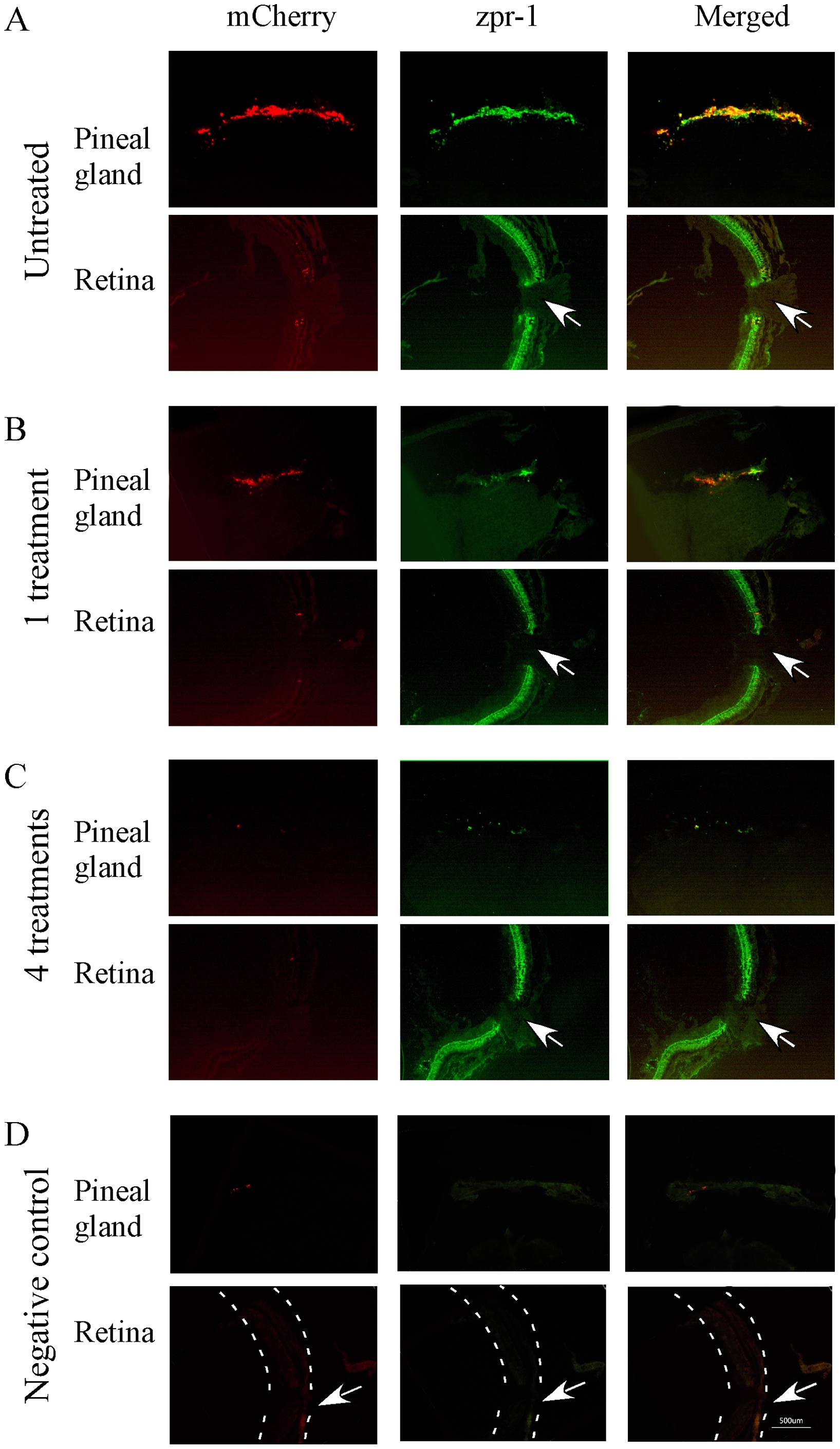
Fig. 4
Cryostat sections of pineal gland and retina of transgenic zebrafish (8-month old) that show mCherry (left panels), double-cone opsin antibody immunoreactivity (zpr-1, middle panels), and co-localization of mCherry and opsin antibody (right panels) before and after the treatment with metronidazole.
(A) Pineal and retinal sections from untreated transgenic fish. Strong mCherry expression was detected in the pineal gland, but not in the retina. zpr-1 immunoreactivity was seen in both pineal and retinal photoreceptor cells. (B) After 1 metronidazole treatment, mCherry expression and zpr-1 immunoreactivity in the pineal gland were decreased. In the retina, strong zpr-1 immunoreactivity was detected. (C) After 4 metronidazole treatments, the expression of mCherry and zpr-1 immunoreactivity in the pineal gland was completely diminished. Strong zpr-1 immunoreactivity was detected in the retina. (D) Fluorescent images of pineal and retinal sections that were processed without primary antibodies (negative controls). Dashed lines outline the retina. Arrows point to the optic nerve.