
|
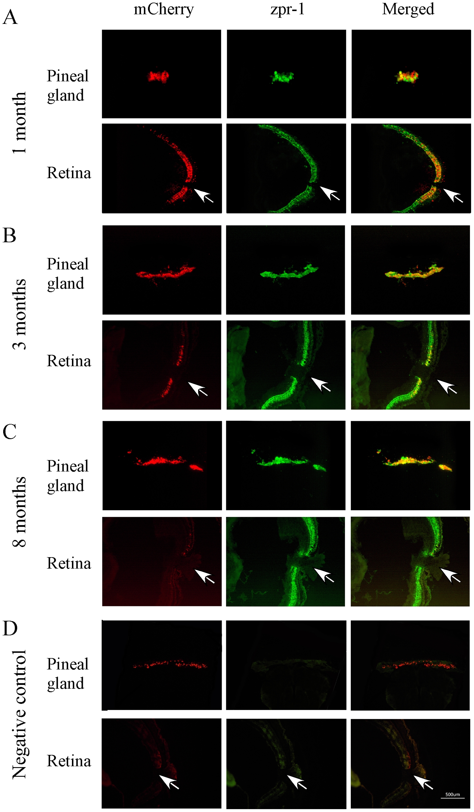
Fig. 3
Cryostat sections of pineal gland and retina of transgenic zebrafish (1, 3 and 8 months) that show mCherry (left panels), double-cone opsin antibody (zpr-1) immunoreactivity (middle panels), and co-localization of mCherry and double-cone opsin antibody (right panels).
(A) At 1 month of age, strong transgene expression was seen in the pineal gland and retina. mCherry and zpr-1 immunoreactivity were co-localized (B) At 3 months, the expression of mCherry decreased in the retina, but increased in the pineal gland. Co-localized of mCherry and zpr-1 immunoreactivity was detected in the pineal gland and central retina. (C) By 8 months, mCherry was detected in the pineal gland, but not in the retina. (D) Fluorescent images of pineal and retinal sections (from 8-months-old animals) that were processed without primary antibodies (negative controls). Arrows point the optic nerve.