FIGURE SUMMARY
Title

MicroRNAs and micromanaging the skeleton in disease, development and evolution

Authors
He, X., Eberhart, J.K., and Postlethwait, J.H.
Source
Full text @ J. Cell. Mol. Med.
1

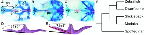
Closely related species and populations within a species often display subtle morphological differences in skeletal systems. Such morphological variation probably results from slight differences in the timing or location or intensity of the action of specific genes. (AC) Skeletal variation in the palate and neurocrania of three species of teleost fish. (A)Danio rerio (zebrafish), (B) Gasterosteus aculeatus (stickleback) and (C) Oryzias latipes (medaka). Note species-specific differences in the shapes of the palate, consisting of ethmoid plate and parasphenoid. (DE) Anterior view of the premaxilla bone showing variation in shape between D. rerio (zebrafish) (D) and D. nigrofasciatus (dwarf danio) (E). The angle of zebrafish premaxilla bone is 81 ± 5° (n= 44) for D. rerio while the angle of D. nigrofasciatus is 74 ± 4° (n= 35). (F) Evolutionary relationships of some fish discussed in this review, including the basally diverging non-teleost Lepisosteus oculatus (spotted gar). Abbreviations: e, eye; ep, ethmoid plate; nt, notochord; ol, otolith; pa, palate; pc, parachordal; ps, parasphenoid and tr, trabeculum.

2

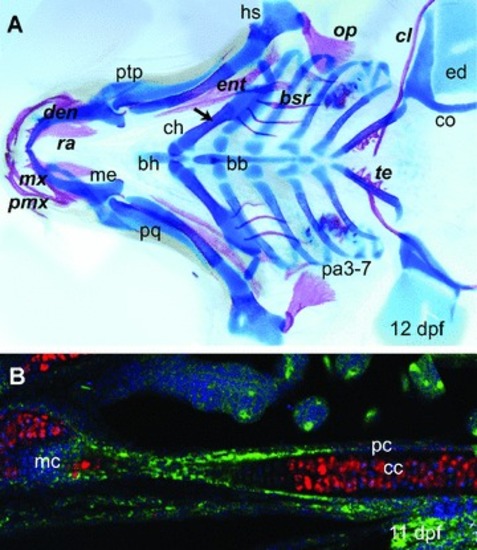
Endochondral ossification and intramembranous ossification. (A) 12-day-old G. aculeatus pharyngeal skeleton stained with Alcian Blue for cartilage and Alizarin Red for bone shows intramembranous and endochondral ossification. Italic bold font labels intramembranous ossification and roman font indicates endochondral ossification. The arrow indicates the endochondral ossification centre of the ceratohyal cartilage. (B) Endochondral ossification of ceratohyal of 11 day old spotted gar. Green, Col II staining includes the perichondrium, which surrounds the cartilage core; red, Col X staining in chondrocytes of the ceratohyal; blue, nuclear stain. Abbreviations: bb, basibranchial; bh, basihyal; bsr, branchiostegal ray; cc, chondrocytes; ch, ceratohyal; cl, cleithrum; co, scapulocoracoid; den, dentary; ed, endoskeletal disc; ent, entopterygoid; hs, hyosymplectic; mx, maxilla; me, Meckel's cartilage; op, opercle; pa, pharyngeal arch; pc, perichondrium, pmx, premaxillary; pq, palatoquadrate; ptp, pterygoid process; ra, retroarticular and te, teeth.

3

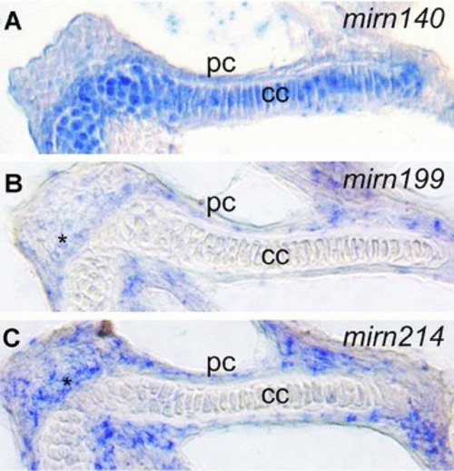
Skeletal miRNAs are expressed in discrete patterns. Conventional in situ hybridization to pre-miRNAs in 3dpf zebrafish larvae shows that mirn140, mirn199 and mirn214 are expressed in the ceratohyal cartilage, confirming results with LNAs [77]. In both skeletal elements, mirn140 is expressed in the chondrocytes while mirn199 and mirn214 are expressed in the perichondrium and surrounding mesenchyme cells. Abbreviations: cc, chondrocytes; mc, mesenchyme and pc, perichondrium.

4

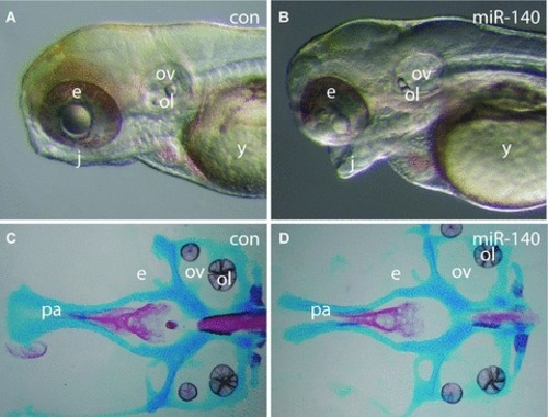
Overexpression of miR-140 causes cleft palate. (A) Whole mount of 3 dpf control zebrafish larva. (B) Whole mount of 3 dpf larva over-expressing miR-140. Note protruding lower jaw. (C) Palate (neurocranium) of control larva. (D) Cleft palate of larva over-expressing miR-140. Abbreviations: con, control; e, eye; j, jaw; miR-140, overexpression construct for miR-140; ol, otolith; ov, otic vesicle; pa, palate and y, yolk.

5

Hox genes are predicted targets for mirn10 and mirn196. Due to genome duplication and subsequent gene loss, zebrafish retains seven clusters of hox genes (gradient grey boxes) that include five copies of mirn196 (blue boxes) and five copies of mirn10 (red boxes). Although protein coding genes of the zebrafish hoxdb cluster were lost, this cluster retained a copy of mirn10[108]. The mirn10 (gradient grey boxes with red surroundings) and mirn196 (gradient grey boxes with blue surroundings) genes both have multiple hox genes as computationally predicted targets.

6

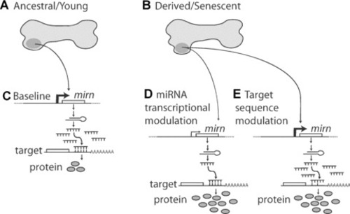
A model for the roles of miRNAs in evolution and disease. (A) A representation of a skeletal element in an evolutionarily ancestral state, or a young or healthy condition. (B) A representation of a skeletal element in an evolutionarily derived state, or a senescent or diseased condition. (C) In the baseline condition, the mirn gene produces a given level of miR molecules, which bind to their target site with a certain affinity to allow a specific level of translation. (D) In the evolutionarily derived state, cis-acting enhancer mutations may decrease or increase (not shown) transcription of the mirn gene, which would lead to decreased (or increased, not shown) inhibition of the target mRNA and hence increased (or decreased, not shown) amounts of target protein, which could alter skeletal shape or function. Similar diminution of mirn expression could occur by stage specific changes in mirn transcription or epigenetic modifications. (E) Mutations accumulated during evolution could alter the miRNA recognition site on target mRNAs to increase or decrease binding, which could alter the amount of target protein produced compared to baseline. Altered protein levels could alter the morphologies or the relative rates of skeletal build-up or degradation by osteoblasts and osteoclasts.

Acknowledgments
This image is the copyrighted work of the attributed author or publisher, and ZFIN has permission only to display this image to its users. Additional permissions should be obtained from the applicable author or publisher of the image. Full text @ J. Cell. Mol. Med.