FIGURE

2

ID
ZDB-FIG-190723-2234
Publication
He et al., 2009 - MicroRNAs and micromanaging the skeleton in disease, development and evolution
Other Figures
All Figure Page
Back to All Figure Page
2

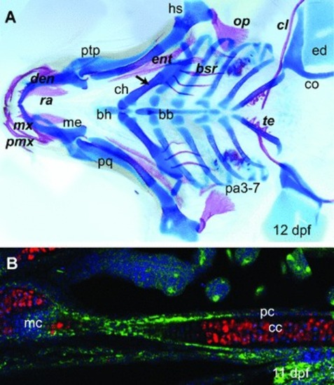
Endochondral ossification and intramembranous ossification. (A) 12-day-old G. aculeatus pharyngeal skeleton stained with Alcian Blue for cartilage and Alizarin Red for bone shows intramembranous and endochondral ossification. Italic bold font labels intramembranous ossification and roman font indicates endochondral ossification. The arrow indicates the endochondral ossification centre of the ceratohyal cartilage. (B) Endochondral ossification of ceratohyal of 11 day old spotted gar. Green, Col II staining includes the perichondrium, which surrounds the cartilage core; red, Col X staining in chondrocytes of the ceratohyal; blue, nuclear stain. Abbreviations: bb, basibranchial; bh, basihyal; bsr, branchiostegal ray; cc, chondrocytes; ch, ceratohyal; cl, cleithrum; co, scapulocoracoid; den, dentary; ed, endoskeletal disc; ent, entopterygoid; hs, hyosymplectic; mx, maxilla; me, Meckel's cartilage; op, opercle; pa, pharyngeal arch; pc, perichondrium, pmx, premaxillary; pq, palatoquadrate; ptp, pterygoid process; ra, retroarticular and te, teeth.

Expression Data

Expression Detail
Antibody Labeling
Phenotype Data

Phenotype Detail
Acknowledgments
This image is the copyrighted work of the attributed author or publisher, and ZFIN has permission only to display this image to its users. Additional permissions should be obtained from the applicable author or publisher of the image. Full text @ J. Cell. Mol. Med.