
- Title
-
Pdlim7 (LMP4) regulation of Tbx5 specifies zebrafish heart atrio-ventricular boundary and valve formation
- Authors
- Camarata, T., Krcmery, J., Snyder, D., Park, S., Topczewski, J., and Simon, H.G.
- Source
- Full text @ Dev. Biol.
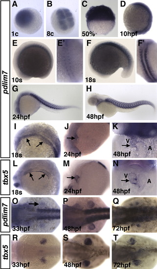
Spatial and temporal expression of zebrafish pdlim7. (A–H) pdlim7 mRNA at (A) 1-cell and (B) 8-cell stages. (C) 50% epiboly. (D) 10 h post-fertilization (hpf). (E) 10-somites. (E′) dorsal view of embryo in E. (F) 18-somites. (F′) dorsal view of embryo in F. Anterior is toward the top in E′ and F′. (G, H) Lateral views of 24 and 48 hpf, respectively. (I–N) pdlim7 (I–K) and tbx5 (L–N) cardiac expression. (I, L) 18-somites stage. (J, M) 24 hpf, (K, N) 48 hpf. Arrows depict expression in cardiac and pectoral fin precursors of the lateral plate mesoderm. (O-T) Expression of pdlim7 (O–Q) and tbx5 (R–T) in the mesenchyme during pectoral fin development. V = ventricle. A = atrium. EXPRESSION / LABELING:
|
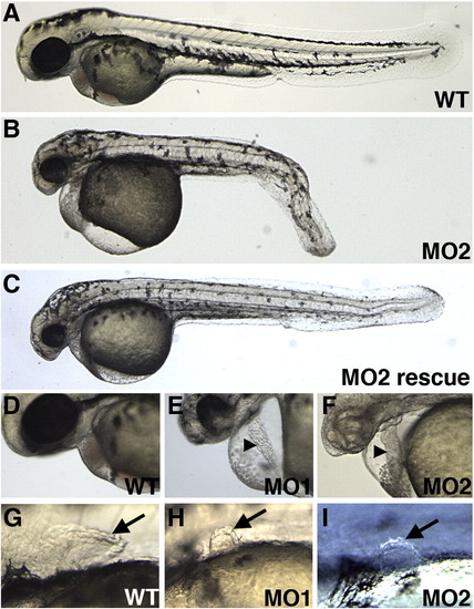
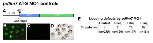
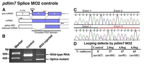
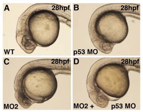
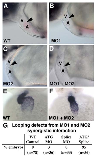
Knock-down of pdlim7 perturbs heart and pectoral fin development. (A–C) Lateral view of wild-type (A), MO2, (B) and rescued MO2 (C) injected embryos at 48 hpf. (D–F) Magnification of hearts of wild-type (D), MO1 (E), and MO2 (F) injected embryo at 48 hpf. Arrowhead in E and F indicates string-like heart. Embryo in F treated with 0.0045% PTU to inhibit pigmentation over the heart. (G–I) Magnification of pectoral fins of wild-type (G), MO1 (H), and MO2 (I) morphants at 48 hpf (arrows). The head is to the left. PHENOTYPE:
|
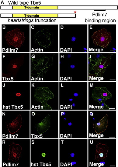
Pdlim7 colocalizes with full-length but not the hearstrings Tbx5. (A) Schematic of wild-type and heartstrings (hst) Tbx5 proteins. Pdlim7 binding region is denoted in wild-type Tbx5. Red asterisk indicates the truncation site of the hst encoded protein. (B–E) Single transfection of myc-Pdlim7 in COS-7 cells. Cells stained with anti-myc antibodies (B), Alexa-488 phalloidin to stain filamentous actin (C), and DAPI nuclear stain (D). (E) Merged image of B–D. (F–I) Single transfection of full-length HA-Tbx5. Cells stained with anti-HA antibodies (F), Alexa-488 phalloidin (G), and DAPI (H). (I) Merged image of F–H. (J–M) Individual transfection of truncated HA-Tbx5 resembling the encoded hst allele. Cells stained for Tbx5 (J), actin (K), and nucleus (L). (M) Merged image of J–L. (N–Q) Cotransfected COS-7 cells stained for Pdlim7 (N), full-length Tbx5 (O), and nucleus (P). (Q) Merged image of N-P. (R-U) Cotransfected cells stained for Pdlim7 (R), truncated Tbx5 (S), and nucleus (T). (U) Merged image of R-T. Scale bar, 20 μm. |
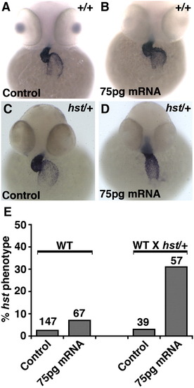
pdlim7 and tbx5 functionally interact during heart development. (A–D) In situ hybridization of 48 hpf embryos with cmlc2. (A) Uninjected wild-type control. (B) Wild-type embryo injected with 75 pg of pdlim7 mRNA. (C) Uninjected hst/+ embryo. (D) hst/+ embryo injected with 75 pg pdlim7 mRNA. (E) Quantification of uninjected and injected embryos with a string-like heart. |
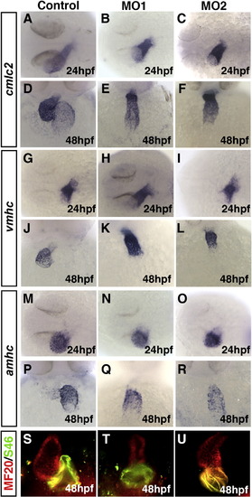
Knock-down of pdlim7 does not alter cardiomyocyte or chamber patterning. (A–F) In situ hybridization of cmlc2 at 24 hpf (A–C) and 48 hpf (D–F) in wild-type (A, D), MO1 (B, E), and MO2 (C, F) injected embryos. (G–L) Expression of vmhc at 24 hpf (G–I) and 48 hpf (J–L) in wild-type (G, J), MO1 (H, K), and MO2 (I, L) injected embryos. (M–S) Expression of amhc at 24 hpf (M–O) and 48 hpf (P–R) in wild-type (M, P), MO1 (N, Q), and MO2 (O, R) injected embryos. (S–U) Frontal view, 48 hpf embryos stained with MF20 and S46 antibodies to detect the ventricle (red) and atrium (yellow) in wild-type (S), MO1 (T), and MO2 (U) injected embryos. |
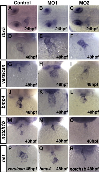
Pdlim7 is required for AV boundary specification. (A–F) In situ hybridization of tbx5 at 24 hpf (A-C) and 48 hpf (D–F) in wild-type (A, D), MO1 (B, E), and MO2 (C, F) injected embryos. (G-O) Expression at 48 hpf of versican (G-I), bmp4 (J-L), and notch1b (M-O). (P-R) Expression of AV boundary markers in hst homozygous embryos. |

Pdlim7 and Tbx5 are required for valve development. (A–D) 72 hpf sagittal sections stained with methylene blue from wild-type (A), MO2 injected (B), and hst embryos (C, D). Black arrowhead indicates myocardium, white arrowhead indicates endocardium in B. Black arrows depict valve tissue in A, C, D. Head is positioned to the right. (E–H) Ventral views of whole mount embryo immunofluorescence at 72 hpf with zn8 (red, Dm-grasp), Alexa 488 phalloidin (green, actin) and DAPI (blue, nuclei) on wild-type (E), MO2 injected (F, G), and hst (H) embryos. White arrows indicate endocardium with Dm-grasp expression in E, F, H. Arrowheads in G denote endocardial layer lacking Dm-grasp. (I–L) Three-dimensional reconstruction of confocal z-stacks at 72 hpf of wild-type (I; Supplemental movie 2), MO2 injected (J, K; Supplemental movies 3 and 4), and hst (L; Supplemental movie 5) embryos. High power views of endocardial Dm-grasp positive cells at the AV boundary (magenta). Myocardial actin (green) is filtered transparent and DAPI nuclei staining has been removed to enhance Dm-grasp visibility (Amira software, see Materials and methods). V = ventricle. A = atrium. Head is positioned to the top. EXPRESSION / LABELING:
PHENOTYPE:
|
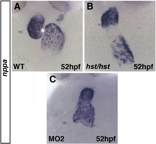
nppa regulation by Tbx5 and Pdlim7. (A-C) In situ hybridization of nppa at 52 hpf in wild-type (A), hst/hst (B), and MO2 injected (C) embryos. Asterisk in A marks AV boundary. |
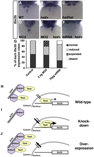
Pdlim7 and Tbx5 directly regulate tbx2b AV expression. (A-C) In situ hybridization of tbx2b 48 hpf at the AV boundary in wild-type (A), hst/+ (B), and hst/hst (C) embryos. (D) AV boundary expansion of tbx2b in wild-type embryo after 2 ng MO2 injection. (E) tbx2b expression in hst/+ embryo injected with MO2 or (F) injected with 75 pg pdlim7 mRNA. (G) Quantification of tbx2b expression levels in uninjected controls (n = 94), 2 ng MO2 (n = 70), and 75 pg mRNA (n = 92). (H-J) Schematics illustrating the effect on the Tbx5 target gene tbx2b in wild-type (H), Pdlim7 MO (I), or pdlim7 mRNA (J) injected embryos. |
PHENOTYPE:
|
PHENOTYPE:
|
PHENOTYPE:
|
PHENOTYPE:
|
Reprinted from Developmental Biology, 337(2), Camarata, T., Krcmery, J., Snyder, D., Park, S., Topczewski, J., and Simon, H.G., Pdlim7 (LMP4) regulation of Tbx5 specifies zebrafish heart atrio-ventricular boundary and valve formation, 233-245, Copyright (2010) with permission from Elsevier. Full text @ Dev. Biol.