
- Title
-
Prdm1a is necessary for posterior pharyngeal arch development in zebrafish
- Authors
- Birkholz, D.A., Killian, E.C., George, K.M., and Artinger, K.B.
- Source
- Full text @ Dev. Dyn.
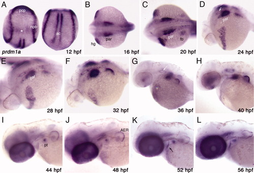
prdm1a expression in the developing zebrafish embryo A: At the beginning of CNCC migration (12 hpf), prdm1a is expressed at the neural plate border (npb), in the adaxial cells (ac), and in the prechordal plate (pcp). It may also be expressed in the otic placode (asterisk). B: By 16 hpf, it is expressed in the pharyngeal arch region (par) and hatching gland (hg). C-E: At 20, 24, and 28 hpf, prdm1a expression continues to progress posteriorly in the arch region (par) and can be detected in the developing pectoral fin bud (fb). F-J: From 32-44 hpf, expression becomes restricted to the more posterior arches and, by 48 hpf, can be detected only in the most posterior arch in the pharyngeal teeth (pt) as well as the AER of the fin bud. K,L: At both 52 and 56 hpf, in addition to the arch and fin bud expression, prdm1a may be expressed in an anterior endodermal pouch (black arrow). (A) dorsal views, anterior to the top; (B,C) dorsal view, anterior to the left; (D-L) lateral view, anterior to the left. White arrows in D and G mark the location of the otic vesicle; ac, adaxial cells; AER, apical ectodermal ridge; fb, fin bud; hg, hatching gland; npb, neural plate border; par, pharyngeal arch region; pt, pharyngeal teeth; pcp, prechordal plate. EXPRESSION / LABELING:
|
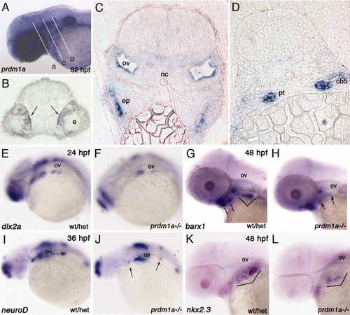
prdm1a is expressed in multiple pharyngeal arch tissues and prdm1a-/- shows a reduction in expression in pharyngeal arch markers. A-D: In situ hybridization showing prdm1a expression in a 52-hpf embryo. A: Whole mount embryo in which the RNA probe was overdeveloped with BM purple and white lines indicate the plane of sectioning in the following panels. B: prdm1a is expressed in the developing neural retina of the eye (black arrows, e). C: The otic vesicle (ov) and an anterior endodermal pouch (ep). D: The developing pharyngeal tooth bud (pt) and mesenchyme of the 5th ceratobranchial (cb5). Molecular markers in the pharyngeal arch region in prdm1a mutants. E, F: dlx2a expression in the post-migratory neural crest in wildtype (E) and prdm1a mutants (F) shows an overall reduction in expression of dlx2a throughout the arch region in prdm1a mutants, but more strongly reduced posteriorly. G, H: barx1 expression in chondrogenic progenitors is slightly reduced in arch 2 and significantly reduced in arches 3-7 in prdm1a mutants (H) as compared to control (G). Bracketed area indicates pharyngeal arch region in wt/het and arrows indicate reduced expression domains in prdm1a-/-. I, J: neuroD expression in neurons of the cranial ganglia shows a reduction of expression in the trigeminal (anterior arrow) and posterior ganglia, including the lateral line ganglia (posterior arrow) in prdm1a mutants (J) as compared to control (I). K,L: nkx2.3 expression in the pharyngeal endoderm of wildtype (K) and prdm1a mutants (L). nkx2.3 is reduced in the posterior arch region in prdm1a mutants. Bracketed area indicates pharyngeal arch region. Anterior is to the left (A, E-L). The otic vesicle (ov) is labeled (E-L) as a reference point. cb5, ceratobrachical 5; ep, endodermal pouch; nc, notochord; ov, otic vesicle; pt, pharyngeal tooth; wt/het, wildtype/heterozygotes embryo. |
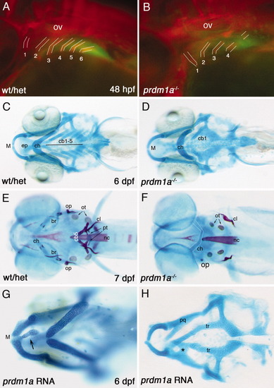
Loss and gain of function of prdm1a results in craniofacial defects. A: 48 hpf prdm1a-GFP embryo stained with zn8 antibody showing the normal structure of the pharyngeal endodermal pouches. B: A prdm1a mutant (in a tg{prdm1a::GFP} background) stained with anti-zn8 shows defects in endodermal pouch patterning. C: A 6-dpf wild type larvae stained with alcian blue to detect cartilage. D: As compared to control, the prdm1a mutant larva has a slightly inverted ceratohyal (ch) and is missing the cartilages of pharyngeal arches 4-7. E: Control 7-dpf zebrafish stained with alcian blue (cartilage) and alizarin red (endochondral and dermal bone). F: As compared to control, the prdm1a mutant larva is missing a majority of the dermal bones, as well as the 5th ceratobranchial and pharyngeal teeth, but the cleithrum is present. The otoliths in the ear are also slightly smaller. The inverted ceratohyal is outlined in white. Staining was performed at multiple timepoints and did not reflect any developmental delay in prdm1a mutants. Overexpression of prdm1a-RNA results in malformed cartilages, including (G) ectopic cartilage extending from Meckel′s cartilage (arrow). H: In addition, a fusion of the pterygoid process with a disarrayed neurocranium is observed. A,B: Lateral views, anterior to the left; C-H: ventral views, anterior is to the left. br, branchiostegal ray; cb1-5, ceratobranchial 1-5; ch, ceratohyal; cl, cleithrum; ep, ethmoid plate; M, Meckel′s cartilage; nc, notochord; op, opercle; ot, otoliths; ov, otic vesicle; pq, palatoquadrate; pt, pharyngeal teeth; tr, trabeculae. 1-6 indicate endodemal pouches; wt/het, wildtype/heterozygotes embryo. |
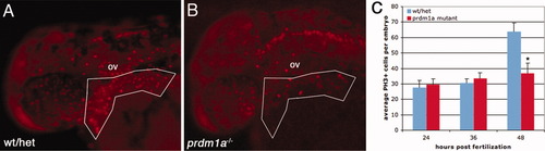
prdm1a is necessary for proliferation within the arches. A: A 48-hpf control embryo stained with anti-phosphohistone H3. B: As compared to control, a 48-hpf prdm1a mutant embryo has a reduction in proliferation in the arch region. C: Average cell proliferation at 24, 36, and 48 hpf in control and prdm1a mutant embryos. Boxed area indicates approximate region in which cell counts were measured and analyzed using Image J and found to not be different in size (not shown). Number of embryos examined for 24 hpf, n = 17; 36 hpf, n = 27; and 48 hpf, n = 13. PHENOTYPE:
|
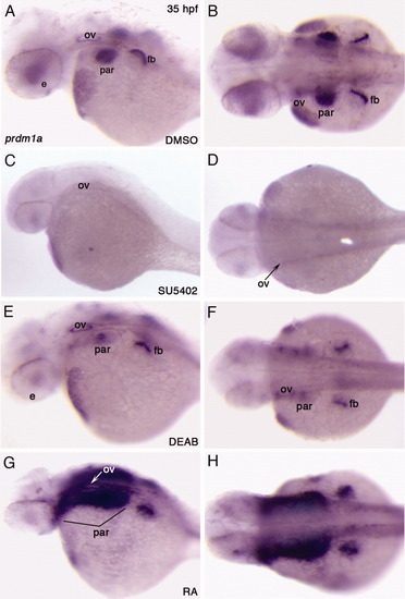
Fgf and RA signaling regulate prdm1a expression in the pharyngeal arches. A,B: Embryos treated with DMSO, as a vehicle control, showing prdm1a expression at 35 hpf. C,D: Treatment with 20 μM SU5402 from 29-35 hpf shows reduced expression of prdm1a in the arches, otic vesicle (arrow), and fin bud. Expression remained in the hatching gland and cloaca (not shown). E,F: Treatment with 50 μM DEAB from 29-35-hpf shows reduced prdm1a expression in the posterior arches (par) and mildly in the fin bud (fb). G,H: Treatment with 0.1 μM RA from 29-35-hpf dramatically increased prdm1a expression in the arches and slightly in the fin bud. prdm1a expansion appears to be endoderm/arch-specific as staining does not cross into the dorsal midline/medial endoderm. All embryos were fixed after treatment and in situ hybridization performed at 35 hpf. A,C,E,G: Lateral views; dorsal to the top, anterior to the left. B,D,F,H: Dorsal views, anterior to the left. e, eye; fb, fin bud; ov, otic vesicle; par, pharyngeal arch region. Bracket in G indicates extent of prdm1a expansion. EXPRESSION / LABELING:
|
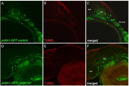
Fluorescent TUNEL staining in prdm1a mutants. A: Lateral image of a 48-hpf control tg{prdm1a::egfp} in the green channel. B: Wildtype/heterozygote (wt/het) embryo labeled with TUNEL (red). C: Merged image of A and B. D: Lateral image of a tg{prdm1a::egfp}; prdm1a mutant embryo. E: Labeled with TUNEL. F: The merged image shows few TUNEL-positive cells and no increase in apoptosis as compared to control. Arrows in B, E show few positive TUNEL cells in both wildtype and prdm1a−/− embryos. |

Unillustrated author statements EXPRESSION / LABELING:
|