
|
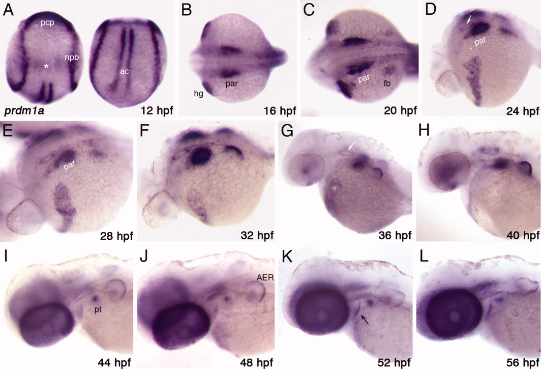
Fig. 1 prdm1a expression in the developing zebrafish embryo A: At the beginning of CNCC migration (12 hpf), prdm1a is expressed at the neural plate border (npb), in the adaxial cells (ac), and in the prechordal plate (pcp). It may also be expressed in the otic placode (asterisk). B: By 16 hpf, it is expressed in the pharyngeal arch region (par) and hatching gland (hg). C-E: At 20, 24, and 28 hpf, prdm1a expression continues to progress posteriorly in the arch region (par) and can be detected in the developing pectoral fin bud (fb). F-J: From 32-44 hpf, expression becomes restricted to the more posterior arches and, by 48 hpf, can be detected only in the most posterior arch in the pharyngeal teeth (pt) as well as the AER of the fin bud. K,L: At both 52 and 56 hpf, in addition to the arch and fin bud expression, prdm1a may be expressed in an anterior endodermal pouch (black arrow). (A) dorsal views, anterior to the top; (B,C) dorsal view, anterior to the left; (D-L) lateral view, anterior to the left. White arrows in D and G mark the location of the otic vesicle; ac, adaxial cells; AER, apical ectodermal ridge; fb, fin bud; hg, hatching gland; npb, neural plate border; par, pharyngeal arch region; pt, pharyngeal teeth; pcp, prechordal plate.