FIGURE SUMMARY
Title

Leukocyte tyrosine kinase functions in pigment cell development

Authors
Lopes, S.S., Yang, X., Müller, J., Carney, T.J., McAdow, A.R., Rauch, G.J., Jacoby, A.S., Hurst, L.D., Delfino-Machín, M., Haffter, P., Geisler, R., Johnson, S.L., Ward, A., and Kelsh, R.N.
Source
Full text @ PLoS Genet.

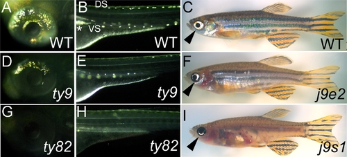
shady mutants form an allelic series with reduced iridophores.
A, B, D, E, G, H) Incident light images of 72 hpf embryos to show eye (A, D, G) and posterior trunk (B, E, H) iridophores (silver spots) in embryonic alleles (D, E, shdty9, weaker; G, H, shdty82, strong) and WT siblings (A, B). C, F, I) Adult viable alleles (F, shdj9e2, weaker; I, shdj9s1, stronger; phenotypic severity shown most clearly by eye iridophores (arrowheads)) and WT sibling (C). In this and subsequent figures, embryos are shown in lateral view, except where noted. DS, dorsal stripe; VS, ventral stripe; *, Lateral patch.

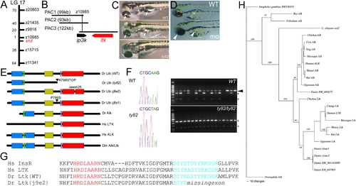
shady mapping and identification as LTK orthologue.
A) shady (shd) map position on LG17; numbers of recombinants in 1000 shd mutant embryos between the marker and shd are given. B) PAC contig in shady region showing gene locations. C) Injection of PAC3 DNA (middle panel) rescues iridophore phenotype (arrows) of shdty82 mutants (no iridophores, lower panel) towards WT (upper panel). D) Injection of ltk morpholino into WT embryo generates shd mutant phenocopies (mo) with much-reduced iridophores (white arrowheads) compared with uninjected sibling (WT) or those injected with control morpholino (not shown). E) Schematics of predicted structures Alk and Ltk proteins in human (Hs), fruit fly (Dm) and zebrafish (Dr). Both WT and 3 mutant variants of the zebrafish are shown. Domains indicated are MAM (blue), LDLa (green), Gly-rich (gold), transmembrane (purple) and tyrosine kinase (red). Proteins are not shown to scale. F) Sequence traces show nucleotide substitution 2415AT (cDNA) in shdty82 (left). RFLP analysis (right) shows homozygosity for the 2415A>T variant (shown by sensitivity to NheI, generating 2 fragments (*)) in 16 shdty82 mutants; 14 WT siblings show only WT allele (NheI insensitive, arrowhead) or are heterozygotes. G) Predicted protein sequence comparison of part of tyrosine kinase domain to show intact catalytic loop (red), but partially deleted activation loop (turquoise), in adult viable shdj9e2 allele due to skipping of exon 26. Sequences are compared with those of human insulin receptor (Hs InsR; A18657) and LTK (Hs LTK; P29376). H) Bayesian analysis of vertebrate ALK/LTK amino acid alignment, using alignment 3 (see Figure S3). Numbers above branches indicate support values for each. Maximum likehood analysis of the same alignment provides the same topology. Translations are of our cDNAs (clones 1 and 3) and other zebrafish genes found by BLAST (XM_686872, XM_001342889 and XM_687805). For accession numbers of other sequences, see Table S2. For details and for other phylogenies obtained, see Figure S3.

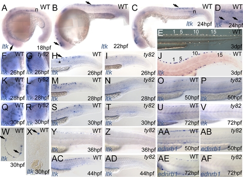
Expression pattern of zebrafish ltk in WT (A–D,F,H,J,K,M,O,Q,S,U,W–Y,AC) and shdty82 homozygous embryos (G,I,L,N,P,R,T,V,Z,AD) throughout embryonic development.
Stages indicated in hpf. A–C) ltk-expressing cells in vicinity of eye (lower arrows in A,B) and in premigratory trunk NC (upper arrow in B and C) and in notochord (n). D) Dorsal view of posterior trunk of WT embryo to show ltk expression in scattered cells in dorsolaterally-positioned subset of premigratory NCCs (arrows). E,J) WT embryo treated with phenylthiourea, illuminated with incident light to show iridophore pattern (E), then fixed and processed for ltk ISH (J); individual cells are numbered. F,G,K,L,Q,R) Dorsoventral spread of ltk-expression in WT eye (F,K,Q); cells remain dorsal to eye in shdty82 mutants (G,L,R). H,I,M,N,S,T,Y,Z,AC,AD) Cells in premigratory (arrow) and migratory (*) positions and in nascent dorsal and ventral stripes are prominent in WT (H,M,S,Y,AC), but almost absent from shdty82 mutants (I,N,T,Z,AD). O,P,U,V,AA,AB,AE,AF) ltk expression pattern closely resembles ednrb1 expression in WT iridophores (AA,AE), but both markers are absent in shdty82 homozygous embryos (P,V,AB,AF). W) Plastic section through eye. X) Transverse section of posterior trunk.

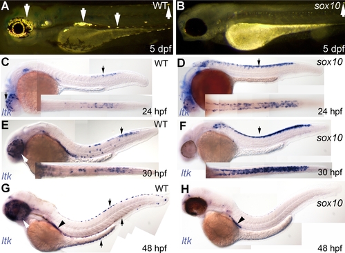
Iridophore phenotype and ltk expression patterns in sox10 mutants.
A, B) Iridophores (arrows) are prominent in 5 dpf WT (A), but almost absent in sox10m618 (1 residual cell is seen here in the dorsal stripe)(B) and sox10t3 (not shown) mutants. C–H) ltk expression (purple, arrows) patterns in WT (C, E, G) and sox10t3 mutants (D, F, H); stages as shown. Arrows indicate ltk-expressing cells on eye and in premigratory NC (24 and 30 hpf) and in iridoblasts (48 hpf); insets show dorsal view of posterior trunk and tail. Arrowhead labels ltk-expressing cells of lateral patch.

EXPRESSION / LABELING:
Gene:
Fish:
Anatomical Terms:
Stage Range: Prim-5 to Long-pec

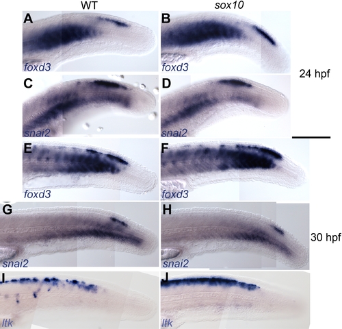
Early NC markers are unaffected in sox10 mutants.
WT (A,C,E,G,I) and sox10t3 mutants (B,D,F,H,J) showing foxd3 (A,B,E,F) or snail2 (C,D,G,H) or ltk expression at 24 (A–D) and 30 hpf (E–J).

EXPRESSION / LABELING:
Genes:
Fish:
Anatomical Term:
Stage: Prim-5

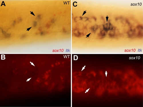
Co-expression of sox10 in ltk-expressing cells in sox10 mutants.
Dorsal views of posterior trunk of 30 hpf sox10m618 (C,D) and WT sibling (A,B) embryos double-labelled for sox10 (red) and ltk (purple, arrows). Autofluorescence from red sox10 signal shown in panels B and D.

EXPRESSION / LABELING:
Genes:
Fish:
Anatomical Term:
Stage: Prim-5

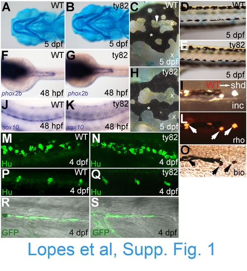
NC derivatives other than iridophores are overtly normal in shd(ty82) mutants. WT siblings (A,C,D,F,J,M,P,R) and shdty82 mutants (B,E,G,H,K,N,Q,S) are shown. A,B) Alcian blue staining of cartilage. C,H) Melanophores (*) and xanthophores (x) in head; iridophores in dorsal head indicated by arrowhead. D,E) Melanophores and xanthophores of posterior trunk. F,G) Enteric nervous system precursors (stained for phox2b mRNA). J,K) Glia of posterior lateral line nerve (sox10). M,N) Enteric neurons of posterior gut (anti-Hu). P,Q) Sensory neurons of tail dorsal root ganglia (anti-Hu immunostaining). R,S) Schwann cells of posterior lateral line nerve (eGFP from 4.9sox10:egfp transgene). Similarly, visual inspection of fin mesenchyme at 4 dpf, anti-Hu immunofluorescence labelling of sympathetic neurons and foxd3-labelled posterior lateral line glia showed no defects in shd mutants (data not shown). I,L,O)Transplants of WT cells into shdty82 mutants rescued iridophore formation (see Table 1). Part of ventral stripe of a 5 dpf WTàshdty82 chimaera to show rescued iridophores (I, incident light). Note that rescued iridophorot shown). I,L,O)Transplants of WT cells into shdty82 mutants rescued iridophore formation (see Table 1). Part of ventral stripe of a 5 des all show both lineage tracers (L, rhodamine dextran; O, biotinylated dextran). Stages as indicated. Embryos are shown in lateral view, except in A and B (ventral view) and C, F-H (dorsal view).

Acknowledgments
This image is the copyrighted work of the attributed author or publisher, and ZFIN has permission only to display this image to its users. Additional permissions should be obtained from the applicable author or publisher of the image. Full text @ PLoS Genet.