shady mapping and identification as LTK orthologue.
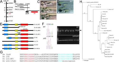
A) shady (shd) map position on LG17; numbers of recombinants in 1000 shd mutant embryos between the marker and shd are given. B) PAC contig in shady region showing gene locations. C) Injection of PAC3 DNA (middle panel) rescues iridophore phenotype (arrows) of shdty82 mutants (no iridophores, lower panel) towards WT (upper panel). D) Injection of ltk morpholino into WT embryo generates shd mutant phenocopies (mo) with much-reduced iridophores (white arrowheads) compared with uninjected sibling (WT) or those injected with control morpholino (not shown). E) Schematics of predicted structures Alk and Ltk proteins in human (Hs), fruit fly (Dm) and zebrafish (Dr). Both WT and 3 mutant variants of the zebrafish are shown. Domains indicated are MAM (blue), LDLa (green), Gly-rich (gold), transmembrane (purple) and tyrosine kinase (red). Proteins are not shown to scale. F) Sequence traces show nucleotide substitution 2415AT (cDNA) in shdty82 (left). RFLP analysis (right) shows homozygosity for the 2415A>T variant (shown by sensitivity to NheI, generating 2 fragments (*)) in 16 shdty82 mutants; 14 WT siblings show only WT allele (NheI insensitive, arrowhead) or are heterozygotes. G) Predicted protein sequence comparison of part of tyrosine kinase domain to show intact catalytic loop (red), but partially deleted activation loop (turquoise), in adult viable shdj9e2 allele due to skipping of exon 26. Sequences are compared with those of human insulin receptor (Hs InsR; A18657) and LTK (Hs LTK; P29376). H) Bayesian analysis of vertebrate ALK/LTK amino acid alignment, using alignment 3 (see Figure S3). Numbers above branches indicate support values for each. Maximum likehood analysis of the same alignment provides the same topology. Translations are of our cDNAs (clones 1 and 3) and other zebrafish genes found by BLAST (XM_686872, XM_001342889 and XM_687805). For accession numbers of other sequences, see Table S2. For details and for other phylogenies obtained, see Figure S3.
|
