
- Title
-
Analysis and functional evaluation of the hair-cell transcriptome
- Authors
- McDermott, B.M., Baucom, J.M., and Hudspeth, A.J.
- Source
- Full text @ Proc. Natl. Acad. Sci. USA
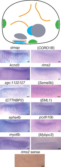
Expression of hair-cell transcripts in the anterior macula. The schematic diagram of the left ear of a 4-day-old zebrafish larva depicts the sensory maculae (blue), cristae of the semicircular canals (green), and otoliths (gray). A red box delimits the anterior macula. In situ hybridization was performed on whole-mount embryos for selected genes identified in Table 1. A bracketed symbol for a human or mouse gene indicates a probe designed to hybridize with a zebrafish transcript for which the corresponding protein′s amino acid sequence most resembles that encoded by the indicated gene. The rims2 sense probe and the myo6 probe constituted, respectively, a negative and a positive control. (Scale bars, 5 μm.) |
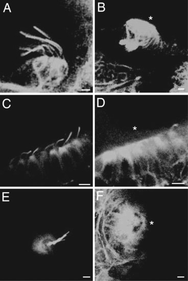
Kinocilia in wild-type and ift172 mutant zebrafish. (A) In a confocal image of the crista from a semicircular canal in a wild-type animal, immunolabeling of acetylated tubulin shows prominent kinocilia. (B) The corresponding image from an ift172 mutant reveals no kinocilia. For this and subsequent instances in which a receptor organ lacks kinocilia, an asterisk indicates the apical hair-cell surface. (C) Numerous kinocilia are apparent in a control preparation from the anterior macula. (D) Although the outlines of hair cells are clearly visible in the anterior macula of a mutant, all of the kinocilia but one are missing. (E) A neuromast from the lateral line of a wild-type animal displays a cluster of kinocilia. (F) The kinocilia are absent from a mutant. (Scale bars, 5 μm.) |
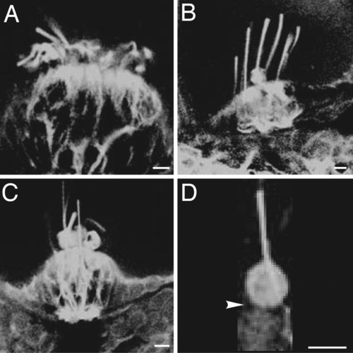
Effect of the seahorse mutation on the kinocilia of 4-day-old zebrafish. (A) Numerous kinocilia, labeled by an antiserum against acetylated α-tubulin, protrude from the crista of semicircular canal in a wild-type animal. (B) The kinocilia of a seahorse mutant larva displays enlarged kinocilia, one with a basal swelling. (C) In another mutant animal, several kinocilia are bloated at their bases. (D) A higher-magnification view of one hair cell from a seahorse larva depicts the swollen base of a kinocilium; the arrowhead indicates the position of the hair cell′s apical surface. (Scale bars, 5μm.) |