
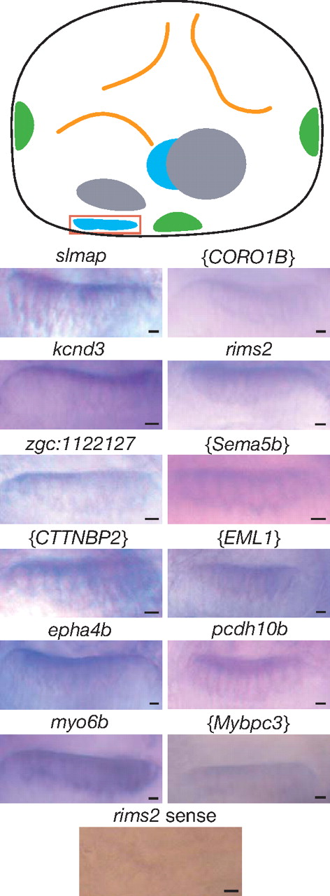
Fig. 1
|
Fig. 1
Expression of hair-cell transcripts in the anterior macula. The schematic diagram of the left ear of a 4-day-old zebrafish larva depicts the sensory maculae (blue), cristae of the semicircular canals (green), and otoliths (gray). A red box delimits the anterior macula. In situ hybridization was performed on whole-mount embryos for selected genes identified in Table 1. A bracketed symbol for a human or mouse gene indicates a probe designed to hybridize with a zebrafish transcript for which the corresponding protein′s amino acid sequence most resembles that encoded by the indicated gene. The rims2 sense probe and the myo6 probe constituted, respectively, a negative and a positive control. (Scale bars, 5 μm.)