
|
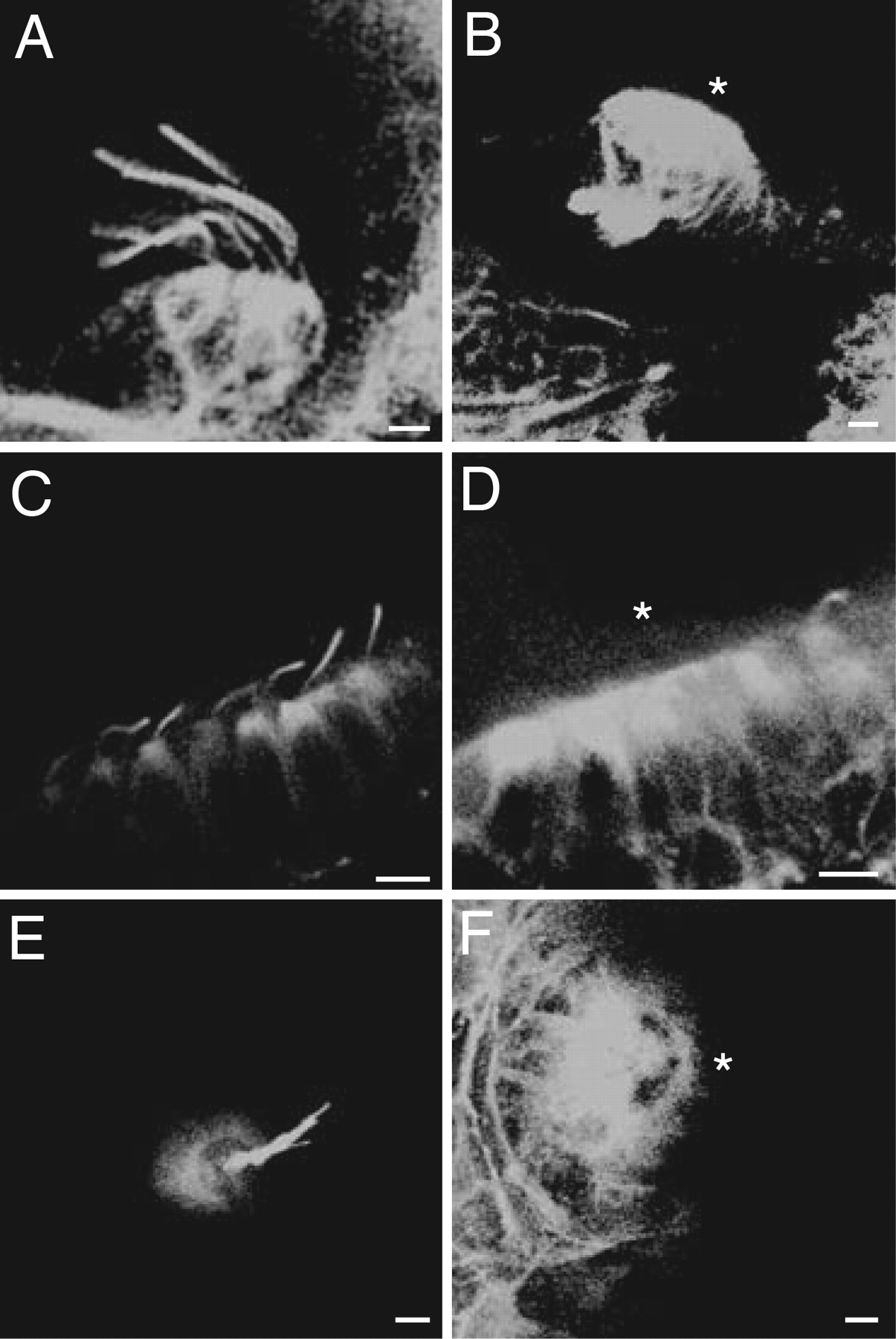
Fig. 2
Kinocilia in wild-type and ift172 mutant zebrafish. (A) In a confocal image of the crista from a semicircular canal in a wild-type animal, immunolabeling of acetylated tubulin shows prominent kinocilia. (B) The corresponding image from an ift172 mutant reveals no kinocilia. For this and subsequent instances in which a receptor organ lacks kinocilia, an asterisk indicates the apical hair-cell surface. (C) Numerous kinocilia are apparent in a control preparation from the anterior macula. (D) Although the outlines of hair cells are clearly visible in the anterior macula of a mutant, all of the kinocilia but one are missing. (E) A neuromast from the lateral line of a wild-type animal displays a cluster of kinocilia. (F) The kinocilia are absent from a mutant. (Scale bars, 5 μm.)