
- Title
-
Novel antizyme gene in Danio rerio expressed in brain and retina
- Authors
- Ivanov, I.P., Pittman, A.J., Chien, C.B., Gesteland, R.F., and Atkins, J.F.
- Source
- Full text @ Gene
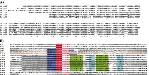
A) Protein alignment of AZR and five other vertebrate antizymes. The positions of amino acids identical in all six proteins are indicated by “*” at the bottom. Abbreviations are as follows: Dr = Dano rerio; Hs = Homo sapiens. B) mRNA alignment of the region surrounding the antizyme frameshift site. The frameshift site is in red letters. The base-pairing blocks of vertebrate RNA pseudoknots are highlighted in green and light-blue (like base-pairing with like). The sequence of AZR is shown in orange letters and the nucleotides 3′ of the frameshift site that display limited nucleotide conservation when compared to other vertebrate and some invertebrate antizymes are highlighted in magenta. The proximal, middle and distal 5′ stimulating modules are highlighted in red, dark blue and grey respectively. The species used in this figure are: N. crassa, S. pombe, C. elegans, A. gambiae, D. melanogaster, D. rerio (zebrafish), X. laevis, M. musculus, and H. sapiens. |
Phylogenetic tree of antizymes based on their amino acid sequences. The tree was generated using the neighbor-joining method. The length of each branch is proportional to the rate of divergence. The position of AZR is indicated in red. The evolutionary tree of the orthologous groups in fish is indicated by magenta coloring of the branches. The species used in this figure are: Saccharomyces cerevisiae, Schizosaccharomyces pombe, Caenorhabditis elegans, Drosophila melanogaster, Danio rerio, Pimephales promelas (minnow), Tetraodon nigroviridis (puffer fish), Gasterosteus aculeatus (three-spined stickleback), Gallus gallus, Xenopus laevis, Mus musculus and Homo sapiens. |
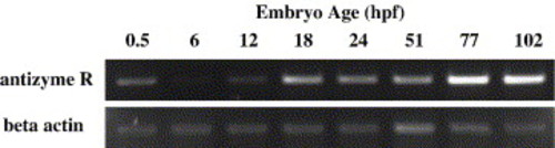
Temporal expression of antizyme from 0.5 hpf to 102 hpf detected by reverse transcription polymerase chain reaction (RT-PCR). Antizyme is maternally expressed (0.5 hpf) and at 6 hpf is only weakly expressed. Antizyme transcript levels increase as development proceeds. Beta-actin was used as a loading control. |
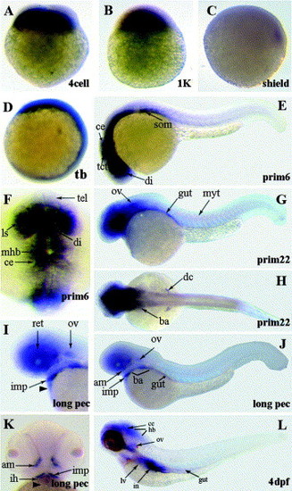
Whole-mount in situ hybridization of AZR at 72 hpf and 5 dpf with sense and anti-sense probes (signal shown as purple). A) Overview shows staining in somites and brain with anti-sense probe (top) but not with sense probe (bottom). Lateral views, dorsal is up, anterior to right. B) Ventral view of the head. AZR mRNA is expressed in the brain and in the retinal ganglion cell (RGC) layer (arrow) of the eye. In this view the RGC layer is a semicircular annulus between the lens and the darkly-staining inner plexiform layer, which contains RGC dendrites. Top, anti-sense probe; bottom, sense control. C) Lateral view of 5 dpf dissected eyes showing expression in the mature retinal ganglion cell layer (arrow), dorsal is up. In this view the RGC layer is an annulus between the lens and the inner plexiform layer. Top, anti-sense probe; bottom, sense control. Scale bars = 100 μm. |
Reprinted from Gene, 387(1-2), Ivanov, I.P., Pittman, A.J., Chien, C.B., Gesteland, R.F., and Atkins, J.F., Novel antizyme gene in Danio rerio expressed in brain and retina, 87-92, Copyright (2007) with permission from Elsevier. Full text @ Gene