
Fig. 1
- ID
- ZDB-FIG-071001-105
- Publication
- Ivanov et al., 2007 - Novel antizyme gene in Danio rerio expressed in brain and retina
- Other Figures
- All Figure Page
- Back to All Figure Page
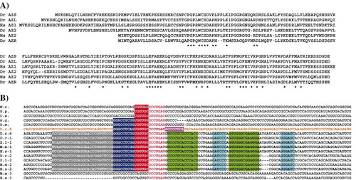
A) Protein alignment of AZR and five other vertebrate antizymes. The positions of amino acids identical in all six proteins are indicated by “*” at the bottom. Abbreviations are as follows: Dr = Dano rerio; Hs = Homo sapiens. B) mRNA alignment of the region surrounding the antizyme frameshift site. The frameshift site is in red letters. The base-pairing blocks of vertebrate RNA pseudoknots are highlighted in green and light-blue (like base-pairing with like). The sequence of AZR is shown in orange letters and the nucleotides 3′ of the frameshift site that display limited nucleotide conservation when compared to other vertebrate and some invertebrate antizymes are highlighted in magenta. The proximal, middle and distal 5′ stimulating modules are highlighted in red, dark blue and grey respectively. The species used in this figure are: N. crassa, S. pombe, C. elegans, A. gambiae, D. melanogaster, D. rerio (zebrafish), X. laevis, M. musculus, and H. sapiens. |
Reprinted from Gene, 387(1-2), Ivanov, I.P., Pittman, A.J., Chien, C.B., Gesteland, R.F., and Atkins, J.F., Novel antizyme gene in Danio rerio expressed in brain and retina, 87-92, Copyright (2007) with permission from Elsevier. Full text @ Gene