
Fig. 4
- ID
- ZDB-FIG-070313-10
- Publication
- Ivanov et al., 2007 - Novel antizyme gene in Danio rerio expressed in brain and retina
- Other Figures
- All Figure Page
- Back to All Figure Page
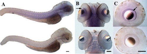
Whole-mount in situ hybridization of AZR at 72 hpf and 5 dpf with sense and anti-sense probes (signal shown as purple). A) Overview shows staining in somites and brain with anti-sense probe (top) but not with sense probe (bottom). Lateral views, dorsal is up, anterior to right. B) Ventral view of the head. AZR mRNA is expressed in the brain and in the retinal ganglion cell (RGC) layer (arrow) of the eye. In this view the RGC layer is a semicircular annulus between the lens and the darkly-staining inner plexiform layer, which contains RGC dendrites. Top, anti-sense probe; bottom, sense control. C) Lateral view of 5 dpf dissected eyes showing expression in the mature retinal ganglion cell layer (arrow), dorsal is up. In this view the RGC layer is an annulus between the lens and the inner plexiform layer. Top, anti-sense probe; bottom, sense control. Scale bars = 100 μm. |
| Gene: | |
|---|---|
| Fish: | |
| Anatomical Terms: | |
| Stage Range: | Pec-fin to Day 5 |
Reprinted from Gene, 387(1-2), Ivanov, I.P., Pittman, A.J., Chien, C.B., Gesteland, R.F., and Atkins, J.F., Novel antizyme gene in Danio rerio expressed in brain and retina, 87-92, Copyright (2007) with permission from Elsevier. Full text @ Gene