
- Title
-
Characterization of dermacan, a novel zebrafish lectican gene, expressed in dermal bones
- Authors
- Kang, J.S., Oohashi, T., Kawakami, Y., Bekku, Y., Izpisua, B.J.C., and Ninomiya, Y.
- Source
- Full text @ Mech. Dev.
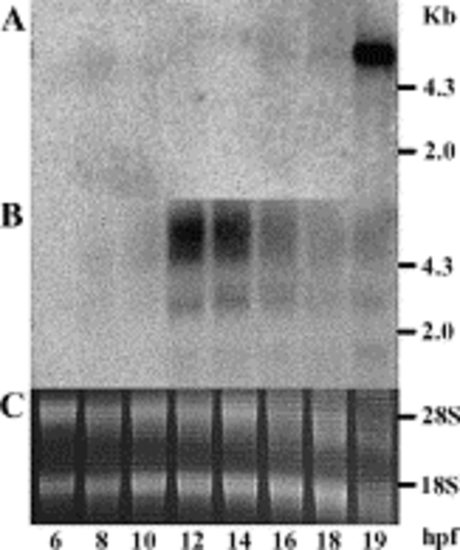
Comparison of expression of dermacan and versican by Northern blot. Total RNA from 6, 8, 10, 12, 14, 16, 18, and 19 hpf was hybridized with a dermacan cDNA (A) and versican cDNA (B) probe specific for the C-terminal globular domain. To show the quality and integrity of RNA, rRNA was visualized by ethidium bromide staining (C). The molecular sizes corresponding to the 28S and 18S ribosomal RNA bands are marked on the right (A,B; according to Schibler et al. (1975)). EXPRESSION / LABELING:
|
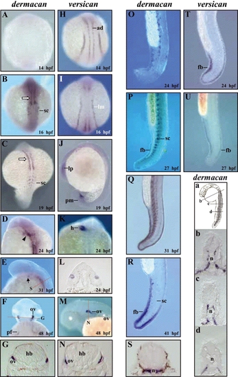
Comparison of embryonic expression of dermacan and versican by whole-mount in situ hybridization. Dorsal views with anterior to the top (A–D,F,H,I,K). Lateral views with anterior to the top (O–R,T,U) and anterior to the left (E,J,M). (A–G) dermacan expression was first detected in the sclerotome and cephalic paraxial mesoderm (open arrow) at 16 hpf (B) and becomes more evident as embryos develop (C, 19 hpf). The expression of dermacan was observed in the cluster of cells between the midbrain and the yolk (arrowhead) at 24 hpf (D), cranial region (arrow) at 31 hpf (E), and otic vesicle and pectoral fin buds at 48 hpf (F). (G) A cross-section of F. (H–N) The expression of versican was observed in adaxial cell and lateral plate mesoderm before dermacan expression (H, 14 hpf, I, 16 hpf), lens primordium and paraxial mesoderm at 19 hpf (J), heart at 24 hpf (K), and otic vesicle at 48 hpf (M). (L) A cross-section in panel J shows the signal in the lens at 24 hpf. (N) A cross-section of M. (O–R) The expression of dermacan in the sclerotome and tail fin bud. dermacan expression was detected in the sclerotome at 24 hpf (O), 27 hpf (P), and 31 hpf (Q), but was not detected at 41 hpf (R). A mesenchymal signal in the tail fin bud was strongly detected at 41 hpf (R). (S) A cross-section of E. versican was detected in tail fin bud at 24 hpf (T) and 27 hpf (U), but was undetectable at 31 hpf (data not shown). (a) Diagram showing the embryo at 23 hpf. (b–d) Cross-sections of embryo in panel a, indicating the signal in the ventral portion of the somites and around the notochord. Abbreviations: ad, adaxial cell; fb, tail fin bud; h, heart; hb, hindbrain; lm, lateral plate mesoderm; lp, lens primordium; n, notochord; ov, otic vesicle; pf, pectoral fin; pm, paraxial mesoderm; sc, sclerotome. EXPRESSION / LABELING:
|
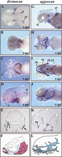
A comparison of the expression pattern of dermacan with that of aggrecan during larval development. Anterior to the left. (A) Dorsal view. (D,G,J) Lateral views. (B,C,H,I) Ventral views. (A–E) The expression of dermacan was observed in the pectoral fin bud and otic vesicle at 3 dpf (A) and dentary at 5 dpf (C,D). The signal in the opercle (arrows) becomes stronger as the embryo develops as shown at 4 dpf (B) and 5 dpf (C,D). (E) A cross-section of C. Note the strong expression in the opercle (arrow). (G–K) aggrecan was expressed in the neurocranium, pharyngeal arch skeleton, and developing pectoral fin at 3 dpf (G), 4 dpf (H), and 5 dpf (I,J). (K) A cross-section of J showing expression in those structures. (F,L) Drawings of dermal bones (F, red color) and cartilage bones (L, blue color) present at larval development. The dark appearance of pigment cells (some indicated by white dots) in the skin and retina is due to pigment, not staining of mRNA. Abbreviations: abc, anterior basicranial commissure; bb, basibranchial; bh, basihyal; cb, ceratobranchials; ch, ceratohyal; d, dentary; ep, ethmoid plate; hs, hyosymplectic; ih, interhyal; iop, interopercle; mc, Meckel′s cartilage; mx, maxilla; op, opercle; ov, otic vesicle; pbc, posterior basicranial commissure; pf, pectoral fin; pop, preopercle; pp, pelvic process; pq, platoquadrate; s, scapulocoracoid; sop, subopercle; t, trabeculae. EXPRESSION / LABELING:
|
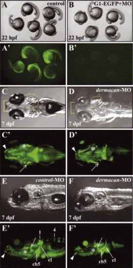
Effects of dermacan morpholino on external morphology and bones at 7 dpf. (A–B′) Downregulation of dermacan G1–EGFP fusion protein translation by dermacan-MO. (A,A′) Control embryos at 22 hpf injected with 20 pg of dermacan G1–EGFP mRNA in the bright field (A) and dark field (A′). Notice the EGFP signals in all embryos (A′). (B, B′) Embryos at 22 hpf co-injected with 20 pg of dermacan G1–EGFP mRNA and 8 ng of dermacan-MO in the bright field (B) and dark field (B2). Note that EGFP signals were completely suppressed in all embryos. (C–F′) dermacan gene targeting by antisense morpholino. (C,C′,E,E′) Embryos injected with control-MO. (D,D′,F,F′) Embryos injected with dermacan-MO. (C′,D′,E′,F′) Bones were fluorescently labeled with Calcein (visualized by green color). (C,C′,E,E′) control-MO injected larvae developed normally. (E′) Vertebrae numbers 1 and 4 were calcified. (D,D′,F,F′) dermacan-MO injected larvae showed several abnormalities. (D,F) Jaw (basket) and gill cover (dotted line) were morphologically defective. (D′,F′) Note that the dentary (arrowheads), opercle (arrows), and branchiostegal ray (asterisks) were either malformed or absent. Vertebrae were not detected in F′. Anterior to the left. (C,C′,D,D′) Ventral views. (E,E′,F,F′) Lateral views. Abbreviations: cb5, ceratobranchial 5; cl, cleithrum; e, eye; ov, otic vesicle; y, yolk. |
Reprinted from Mechanisms of Development, 121(3), Kang, J.S., Oohashi, T., Kawakami, Y., Bekku, Y., Izpisua, B.J.C., and Ninomiya, Y., Characterization of dermacan, a novel zebrafish lectican gene, expressed in dermal bones, 301-312, Copyright (2004) with permission from Elsevier. Full text @ Mech. Dev.