
- Title
-
Vascular endothelial growth factor receptor signaling is required for cardiac valve formation in zebrafish
- Authors
- Lee, Y.M., Cope, J.J., Ackermann, G.E., Goishi, K., Armstrong, E.J., Paw, B.H., and Bischoff, J.
- Source
- Full text @ Dev. Dyn.
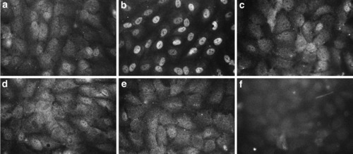
VEGF-induced NFATc1 nuclear translocation was blocked by the VEGF-R tyrosine kinase inhibitors. HPVEC grown for 24 hr in Endothelial Basal Media media with 10% FBS were either mock stimulated (a) or stimulated with 50 ng/ml VEGF165 (R&D Systems) for 30 min (b-f). Intracellular localization of NFATc1 was visualized using an anti-human NFATc1-specific mAb (a-e). NFATc1 expression in untreated HPVECs (a) was detected in the cytoplasm, as evident by the diffuse staining throughout the cells. Nuclear localization of NFATc1 was evident in HPVECs treated with VEGF165 (b). Pretreatment of HPVEC with 1 μM FK506 (c), 2 μM AAC789 (d), or 2 μM PTK787 (e) inhibited VEGF-induced nuclear translocation of NFATc1. VEGF-treated HPVEC incubated with isotype-matched control IgG (f). |
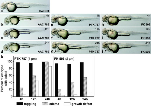
Morphology of AAC 789-, PTK787-, and FK506-treated zebrafish embryos at 48 hpf. Zebrafish embryos staged at 15 somites (17 hpf) were treated with AAC 789 (5 μM), PTK 787 (5 μM), or FK506 (2 μM) for 4, 12, and 24 hr. At the end of each time period, embryos were washed several times with fresh medium to remove the inhibitor drugs and allowed to develop until 48hpf. Treatment for 4 hr with AAC 789 (b), PTK787 (e), or FK506 (h) resulted in toggling of blood within the heart (see Supplementary Movies), compared to control embryos, while no apparent effect on gross morphogenesis was visible. Treatment with PTK 787 for 12 hr induced minor epicardial edema as well as minor retarded craniofacial development (f and k). Treatment with PTK787 for 24 hr resulted in severe epicardial edema (g, arrow), shortened tails, retarded brain growth, and blood toggling in the heart (k). Embryos treated with AAC 789 or FK506 for 12 hr (c, i) and 24 hr (d, j) showed almost the same morphology at 48 hpf as control embryos (a), with the exception of minor epicardial edema in embryos treated for 24 hr with FK506 (j, arrow). k: The number of embryos with defects in each condition and at each time point (n = 160-500) is shown. No embryos in the untreated group showed any regurgitation of blood (n = 178) (not shown). |
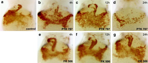
Effects of PTK 787 and FK506 on hemoglobinized blood levels and circulation. Embryos treated with PTK 787 and FK506 as described in Figure 2 were stained with o-dianisidine for circulating hemoglobinized red blood cells (Iuchi and Yamamoto, [1983]) in the yolk sac and heart (30). a: Control embryos at 48 hpf. b: PTK787 treatment for 4 hr had no effect on oxygenated blood levels and circulation. Twelve-hour (c) and 24-hr (d) treatment with PTK 787 resulted in decreased blood in yolk-sac and heart, especially in embryos treated for 24 hr (d). FK506 did not affect oxygenated blood levels or circulation after 4-hr, 12-hr, or 24-hr exposure (panels e, f, g, respectively). |
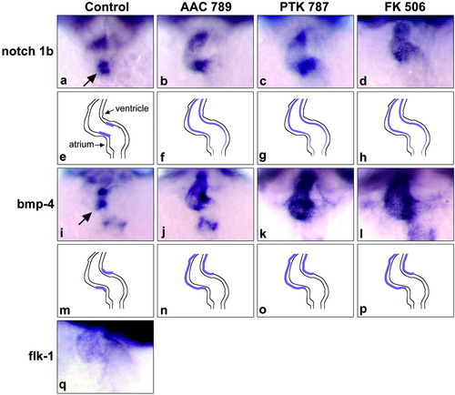
Disruption of VEGF or calcineurin signaling results in altered expression of notch 1b and bmp4 in cardiac valves. In situ hybridization analysis of the endocardial marker notch 1b (a-d), the myocardial marker bmp-4 (i-l), and the endothelial marker flk-1 (q) in the AV valve region. a: notch 1b was restricted to the AV region of the endocardium in control embryos (see arrow). b: notch 1b was ectopically expressed beyond the AV boundary into the ventricle and weakly in the atrium of embryos treated with AAC 789. c,d: Similar ectopic expression of notch 1b in embryos treated with PTK 787 and FK506, respectively. e-h: Schematic figures for the expression patterns seen in a-d. i: bmp-4 expression was prominent in the in myocardial region of AV valve at 48 hpf in control embryos (see arrow). j: AAC 789 treatment for 4 hr resulted in strong ectopic expression of bmp-4, especially in the ventricle region. k,l: PTK787 and FK 506 induced similar mis-localized expression patterns. m-p: Schematic figures for expression patterns of bmp-4 seen i-l. q: Expression of flk-1 in the endocardium of control embryos at 48 hpf. EXPRESSION / LABELING:
|
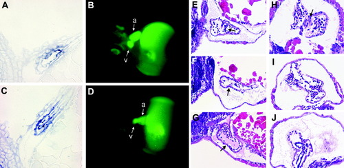
Defects in the AV boundary region of AAC 789-treated embryos. Control embryos (A, B) and embryos treated with 5 μM AAC 789 for 4 hr beginning 17 hpf (C, D) were analyzed by two methods. In situ hybridization for notch 1b was performed on whole mount embryos, which were then embedded in plastic resin, sectioned, and stained with eosin (A,C). Microangiography using FITC-Dextran was performed on embryos at 56 hpf (B, D). Fluorescence images were captured within 30 min of injection. a, atrium; v, ventricle. E-J: H&E stained histological sections of embryos untreated (E,H) or treated with 5 μM PTK 787 for 4 hr beginning at 17 hpf (F, I) or with 2 μM FK (G, J). Sagittal (E-G) or transverse (H-J) sections through embryos at 68 hpf are shown. EXPRESSION / LABELING:
|