
|
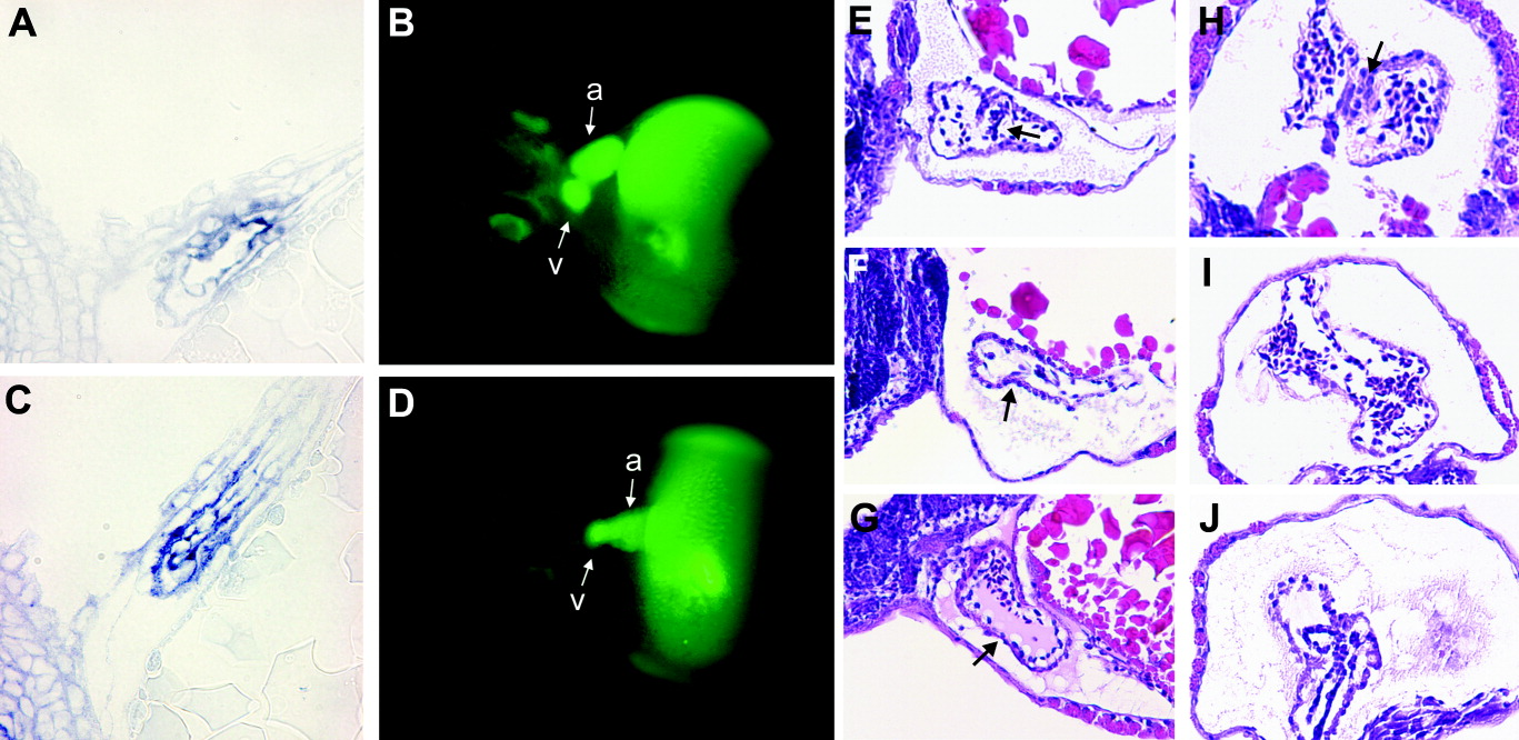
Fig. 5 Defects in the AV boundary region of AAC 789-treated embryos. Control embryos (A, B) and embryos treated with 5 μM AAC 789 for 4 hr beginning 17 hpf (C, D) were analyzed by two methods. In situ hybridization for notch 1b was performed on whole mount embryos, which were then embedded in plastic resin, sectioned, and stained with eosin (A,C). Microangiography using FITC-Dextran was performed on embryos at 56 hpf (B, D). Fluorescence images were captured within 30 min of injection. a, atrium; v, ventricle. E-J: H&E stained histological sections of embryos untreated (E,H) or treated with 5 μM PTK 787 for 4 hr beginning at 17 hpf (F, I) or with 2 μM FK (G, J). Sagittal (E-G) or transverse (H-J) sections through embryos at 68 hpf are shown.