
IMAGE
Fig. 7
- ID
- ZDB-IMAGE-250602-27
- Publication
- Tian et al., 2025 - C-mannosyltransferase DPY19L1L-mediated Reissner Fiber formation is critical for zebrafish (Danio rerio) body axis straightening
- All Figures
- Figures for Tian et al., 2025
Image
|
Figure Caption
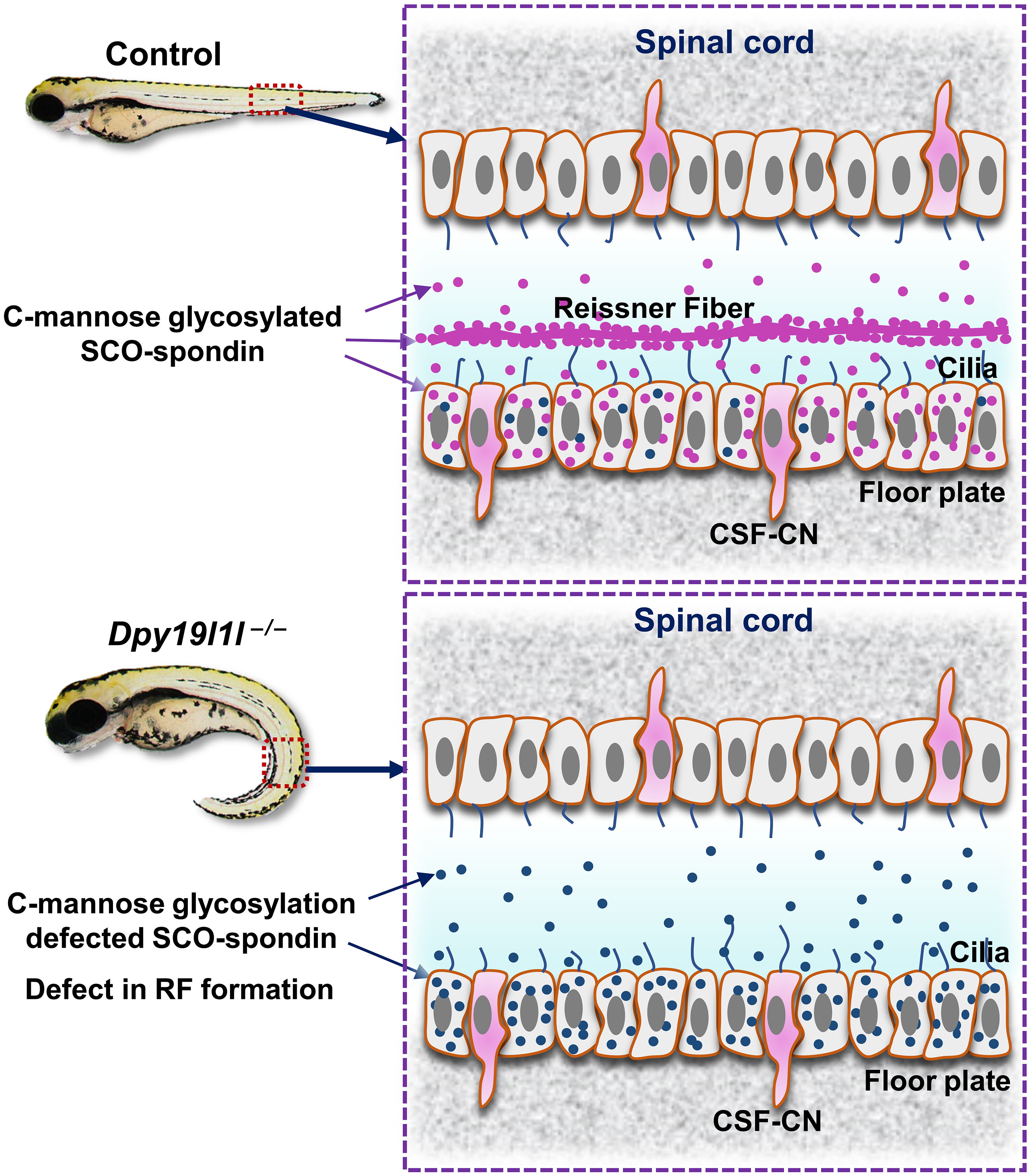
Fig. 7 Model of SCO-spondin and RF in control and dpy19l1l-deficient zebrafish. (Top) In wild-type spinal cord, DPY19L1L C-mannosylates SCO-spondin (purple), enabling RF formation via cilia-driven CSF flow. (Bottom) In dpy19l1l−/− mutants, nonglycosylated SCO-spondin (dark blue) fails to form RF, leading to body axis curvature. CSF-CN, CSF-contacting neuron.
Acknowledgments
This image is the copyrighted work of the attributed author or publisher, and
ZFIN has permission only to display this image to its users.
Additional permissions should be obtained from the applicable author or publisher of the image.
Full text @ Sci Adv