
Image
|
Figure Caption
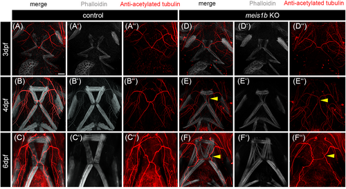
Fig. 9 The position of the trigeminal nerve reflected misshapen muscles. Whole-mount antibody co-staining of nerves with anti-acetylated tubulin (red, A″–C″, D″–F″) and muscles with phalloidin (white, A′–C′, D′–F′). Arrowheads in (E–E″) indicate misshapen pattern of the trigeminal nerve which corresponds to disrupted intermandibularis posterior and hyohyal muscles. Malformation of the trigeminal nerve is even more apparent at 6 dpf in meis1b mutants (arrowheads in F–F″). Scale bar = 30 μm.
Acknowledgments
This image is the copyrighted work of the attributed author or publisher, and
ZFIN has permission only to display this image to its users.
Additional permissions should be obtained from the applicable author or publisher of the image.
Full text @ Dev. Dyn.