
|
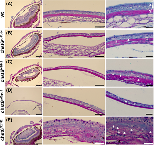
Fig. 7 Histopathology of the corneal tissue in the mutant zebrafish. Alcian blue/periodic acid–Schiff-stained eye sections of (A) wt, (B) chst6pd4/pd4, (C) chst6f42/f42, (D) chst6pd5/pd5, and (E) chst6f42/f42 are shown (n = 7). Left column: 10× overview, scale bars: 200 μm; middle column: 40× close-up overview, scale bars: 50 μm; right column: 100× zoom, scale bars: 20 μm. Serial sections were taken from each eye, and different slices were shown in the middle and right columns. Collagen in stroma is stained pink with periodic acid–Schiff, mucopolysaccharides are stained blue with alcian blue, and nucleus is stained light purple with hematoxylin. e, epithelium; s, stroma. White arrows indicate alcian blue-positive aggregates.