
|
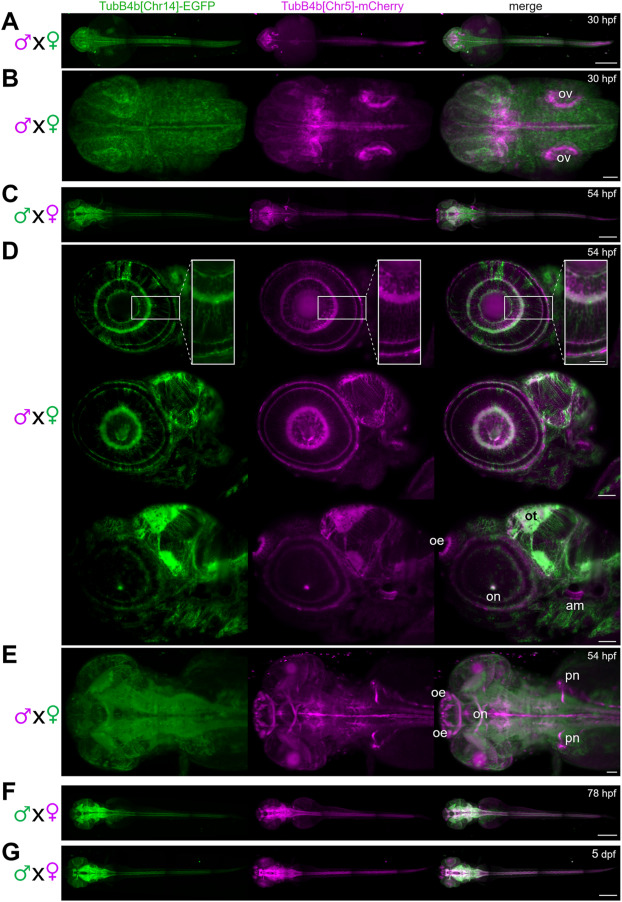
Fig. 4 Expression of TubB4b[Chr14] and TubB4b[Chr5] in developing zebrafish larva. A,B,D,E: double transgenic larva obtained from females Tg(tubB4b[Chr14]:TubB4b[Chr14]-EGFP)+/0 crossed with males Tg(tubB4b[Chr5]:TubB4b[Chr5]-mCherry)+/0 and observed at 30 hpf (A,B) or 54 hpf (D,E). C,F,G: double transgenic larva obtained from females Tg(tubB4b[Chr5]:TubB4b[Chr5]-mCherry)+/0 crossed with males Tg(tubB4b[Chr14]:TubB4b[Chr14]-EGFP)+/0 and observed at 54 hpf (C), 78 hpf (F) or 5 dpf (G). Zeiss Lighsheet imaging of whole larva (A,C,F,G; scale bar = 300 μm) or head region (B,D,E; scale bar = 50 μm; scale bar = 20 μm for inset in D). Dorsal view (A,B,C,E,F,G) or lateral view (D). Projection of stacks in A,B,C,E,F,G; single digital sections in D, from lateral (top panel) to medial (bottom panel). ov: otic vesicle; oe: olfactory epithelium; ot: optic tectum; on: optic nerve; am: anterior macula; pn: pronephros.
Reprinted from Developmental Biology, 517, Smaili, W., Pezet, C., Marlin, S., Ernest, S., R391 human dominant mutation does not affect TubB4b localization and sensory hair cells structure in zebrafish inner ear and lateral line, 301-316, Copyright (2024) with permission from Elsevier. Full text @ Dev. Biol.