
|
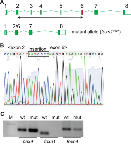
Fig. 1 Characterization of a foxn1 null allele in zebrafish. (A) Schematic structure of the zebrafish foxn1 gene; exons are numbered, and the exons encoding the DNA binding domain are indicated as red rectangles. A double-headed arrow below the schematic indicates the region deleted in the foxn1fr107 mutant allele. (B) Partial sequence of the cDNA spanning the deletion from within exon 2 to within exon 6; note the insertion of seven nucleotides as a result of the CRISPR/Cas9-mediated rearrangement that fuses parts of exons 2 and 6. (C) RT-PCR analysis of the indicated epithelial genes in the thymi of wildtype (wt) and foxn1-deficient (mut) zebrafish (M; marker lane; amplicon sizes: pax9, 149 bp; foxn1, 110 bp; foxn4, 174 bp. Note the absence of an intact foxn1 transcript in the mutant thymus.