
Fig. 1
- ID
- ZDB-IMAGE-240206-26
- Publication
- Birdal et al., 2023 - Expression of taste sentinels, T1R, T2R, and PLCß2, on the passageway for olfactory signals in zebrafish
- All Figures
- Figures for Birdal et al., 2023
|
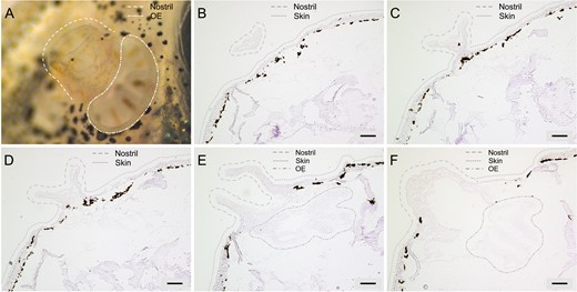
Fig. 1 The funnel-like structure of the zebrafish nostril and its alignment with the OE. The topology of the zebrafish nostril is shown in a photograph and representative coronal sections. The nostril is usually present on 30–40 sections, of which five 10 µm cryostat sections are shown from anteriormost to posteriormost. (A) Photograph of zebrafish nostril taken under dissection microscope (dashed lines), the OE (enclosed in dash-dotted lines) is partially visible below the nasal outlet. (B) Appearance of the coronal sections of the nostrils (enclosed in dashed lines) at the most anterior positions where the connection of the nostril to the head skin (indicated with dotted lines) is not visible yet. (C) The widening of the opening of the nostril and the connection of the nostril to the head skin is observed at more posterior sections. (D) Further widening of the opening of the nostril and the connection of the nostril to the head skin is observed at further posterior sections. (E) The opening of the nostril to the OE is observed in sections where both the nostril and the OE (enclosed in dash-dotted lines) are visible. (F) Coronal sections of the nostrils and the OE at the most posterior positions show the closed parts of the nostril, where water is funneled through contacting the OE before it seeps out of the posterior opening (posterior nostril). Scale bars are 100 µm.