
Image
|
Figure Caption
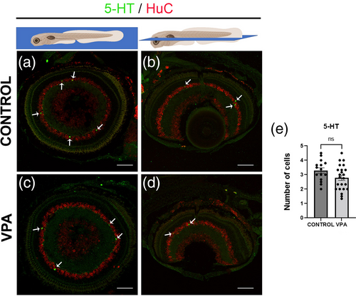
Fig. 3 Serotonergic neurons in the retina of larvae zebrafish at 8-dpf. (a, d) Immunostaining for 5-HT (green) in sagittal and horizontal sections of larval (8-dpf) retinae. 5-HT positive cells (green) are seen in the inner nuclear layer of the retina in both VPA-treated and control larvae in a similar distribution. (e) Quantification of the number of cells per section showed no statistical differences between treated animals and controls. Neurons were counterstained with HuD/HuC label (red). Data are expressed as mean ± SEM, n = 5 per group, *p-value <0.05. 5-HT, serotonin. Scale bar 50 μm.
Acknowledgments
This image is the copyrighted work of the attributed author or publisher, and
ZFIN has permission only to display this image to its users.
Additional permissions should be obtained from the applicable author or publisher of the image.
Full text @ Autism Res