Image
Figure Caption
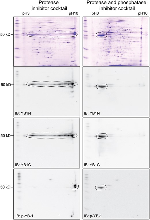
Fig. 5 Western blot evidence for multi-site phosphorylation of Ybx1. The protein extract from PG follicles in the presence or absence of phosphatase inhibitors followed by 2D electrophoresis and Western blot analysis with YB1N, YB1C, and anti-p-YB-1. In the absence of phosphatase inhibitor, the signal of Ybx1 protein spanned a wide range of pH from 3 to 10. This pattern disappeared in the presence of phosphatase inhibitors and the signal became concentrated around pH 3. The anti-p-YB-1 detected phosphorylation at S82 site.
Acknowledgments
This image is the copyrighted work of the attributed author or publisher, and
ZFIN has permission only to display this image to its users.
Additional permissions should be obtained from the applicable author or publisher of the image.
Full text @ Biol. Reprod.

