
Fig. 4
- ID
- ZDB-IMAGE-231229-10
- Genes
- Publication
- Nikaido et al., 2023 - Intestinal expression patterns of transcription factors and markers for interstitial cells in the larval zebrafish
- All Figures
- Figures for Nikaido et al., 2023
|
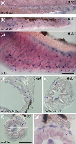
Fig. 4 The intestinal expression of pdgfra gene at different stages. (a–c) Expression of pdgfra gene in the middle and distal part of the intestine at 3 dpf (a) and 6 dpf (b, c). Panel c shows its expression in the intestinal bulb. Skin is removed in the 6 dpf larvae for better observation. Purple signals are observed in the intestine (purple triangles). Left side views. Anterior, to the left. Stages examined are shown in the top-right corners. (d–g) Cross sections at the level of anterior part of the intestinal bulb (d), posterior part of the intestinal bulb (e), and middle part of the intestine (f). Panel g is a close-up image of the indicated area in panel f. Purple triangles indicate expression of pdgfra outside of the mucosal layer. Scale bar: 50 μm.