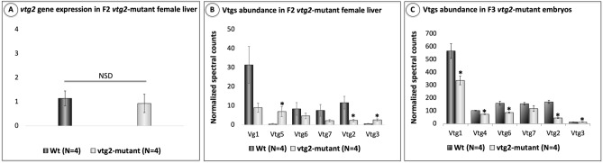
IMAGE

Figure 4

ID
ZDB-IMAGE-231105-5
Source
Figures for Yilmaz et al., 2023
Image
Figure Caption

Figure 4

Relative quantification of vtg2 gene expression in F2 vtg2-mutant zebrafish female liver and Vtg2 protein abundance in 1 hpf F3 vtg2-mutant embryos. (A) Comparison of vtg2 gene expression levels in F2 vtg2-mutant (Ht; vtg2 −/+) and wild type (Wt) female liver in dark and light gray vertical bars, respectively. Vertical brackets indicate SEM. SYBR Green qPCR-2−ΔΔCT mean relative quantification of gene expression normalized to the geometric mean expression of zebrafish elongation factor 1a (eif1a) and ribosomal protein L13a (rpl13a) was employed. Data were statistically analyzed using a Kruskal Wallis nonparametric test p < 0.05. (B) Relative quantification of multiple vitellogenins by LC–MS/MS in F2 vtg2-mutant female (Ht; vtg2 −/+) liver, and (C) Relative quantification of multiple vitellogenins by LC–MS/MS in 1 hpf F3 vtg2-mutant embryos. Comparisons of mean normalized spectral counts for Vtg protein levels in Wt versus vtg2-mutants indicated by dark and light gray vertical bars, respectively. Vertical brackets indicate SEM. Statistically significant differences between group means detected by independent samples Kruskal Wallis non-parametric test (p < 0.05) are indicated by asterisks above bars.

Acknowledgments
This image is the copyrighted work of the attributed author or publisher, and ZFIN has permission only to display this image to its users. Additional permissions should be obtained from the applicable author or publisher of the image. Full text @ Sci. Rep.