Figure Caption
Fig 1
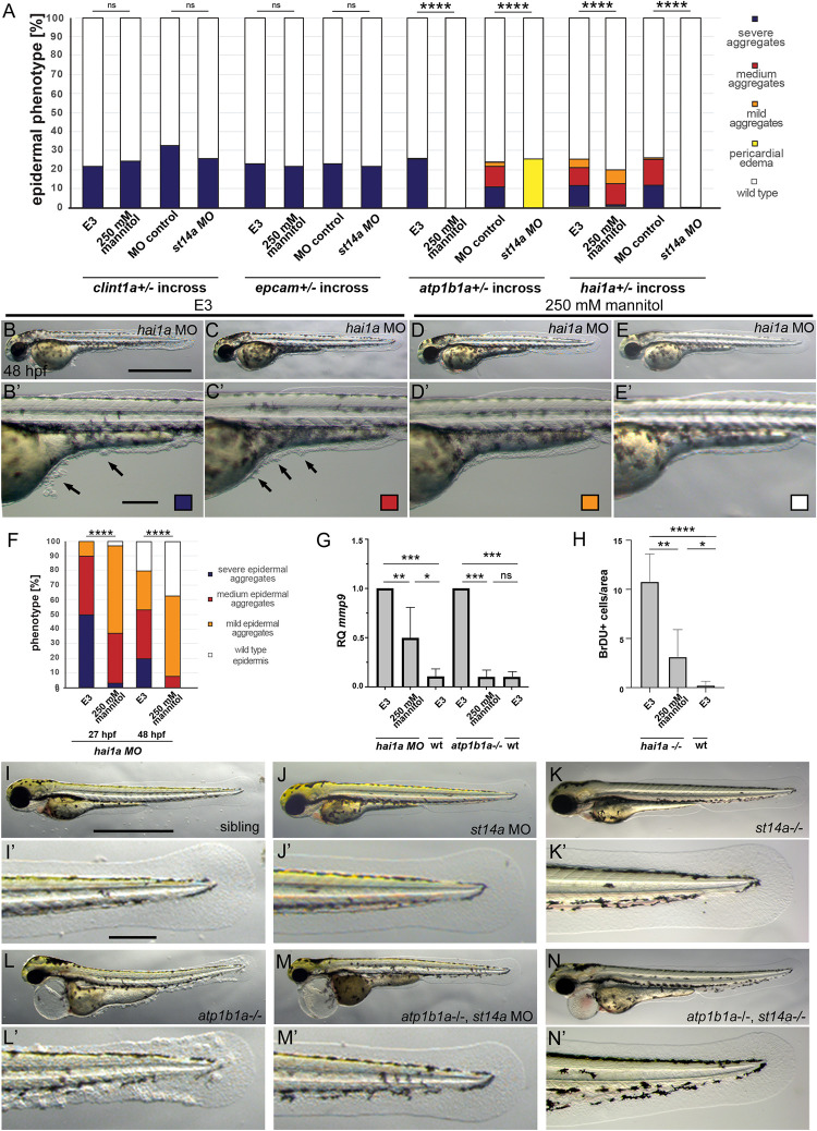
Hypotonic stress and Matriptase-1a function are required for the epidermal phenotype of hai1a and atp1b1a, but not clint1 and epcam mutants.
(A-H) Isotonic medium attenuates the phenotype of hai1a-/- mutants and rescues the phenotype of atp1b1a-/- mutants, but has no effect of clint1-/- or epcam-/- mutants. (A) Quantification of epidermal phenotypes of 48 hpf embryos obtained from parents heterozygous for the mutations clint1hi1520 [22], epcamhi2151 [42], atp1b1am14 [26], or hai1ahi2217 [21], raised in E3, E3 + 250 mM Mannitol, or injected with control morpholino or st14a morpholino (n = 59–102 from N = 3 independent clutches per condition, Significances were determined via a Chi-square test, ns, not significantly different (p>0.05); ****, significantly different (p< 0.0001)). (B-E’) Brightfield images of live 48 hpf hai1a morphants raised in E3 (B,B’,C,C’) or E3 + 250 mM Mannitol (D,D’,E,E’) as lateral overviews of entire embroys (A-D) or magnified lateral views of the yolk tube and yolk extension regions of the same embryos (A’-D’); arrows in (B’,C’) point to epidermal aggregates on yolk sac and ventral median fin fold. (F) Quantification of phenotypic classes (n = 27–67) of hai1a morphant embryos raised in E3 or E3 + 250 mM Mannitol, representatives of which are shown in panels (B-E’) Significances were determined via a Chi-square test, ns, not significantly different (p>0.05); ****, significantly different (p< 0.0001). (G) RT-qPCR showing relative quantities of mmp9 transcript of 48 hpf hai1a morphant or 56 hpf atp1b1a mutant embryos raised in E3 or E3 + 250 mM Mannitol, compared to their respective siblings. cDNA was obtained from pools of 15 embryos each, N = 3 for hai1a, N = 3 for atp1b1a. H. Quantification of BrdU-labeled nuclei in defined, equally-sized areas of the fin fold of wild types and hai1a-/- mutants at 48 hpf, raised in E3 or E3 + 250 mM Mannitol, n = 4–6. Significances in G and H were determined via a one-way ANOVA and Tukey’s post hoc test; ns, not significantly different (p>0.05); *,**,***,****, significantly different (p<0.05, 0.01, 0.001, 0.0001, respectively). (A,I–N’) Loss of st14a function rescues epidermal aggregate formation in atp1b1a-/- mutants. Brightfield images of representative live 72 hpf embryos, either as lateral overviews of the entire embryos (I-N), or as magnified lateral views of the tail of the same embryos (I’-N’): wild-type sibling (I,I’), wild-type sibling injected with st14a MO (J,J’), st14a-/- mutant (K,K’), atp1b1a -/- mutant with epidermal aggregates (L,L’), atp1b1a -/- mutant injected with st14a MO (M,M’), and atp1b1a-/-; st14a-/- double mutant (N,N’), both with wild-type-appearing epidermis. For quantifications, see (A) and S1I Fig. Scale bars: 500 μm (B,I), 100 μm (B’,I’).
Acknowledgments
This image is the copyrighted work of the attributed author or publisher, and
ZFIN has permission only to display this image to its users.
Additional permissions should be obtained from the applicable author or publisher of the image.
Full text @ PLoS Genet.