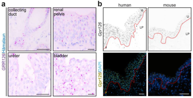
Gpr126 is expressed throughout the urinary collecting system. (a) RNAscope® probes targeting the human GPR126 mRNA were used on human FFPE urinary tissues. GPR126 signal (magenta dots) was found in the epithelial compartment of the urinary collecting system, collecting duct epithelial cells and transitional epithelium (urothelium) of the renal pelvis, ureter, and bladder. Hematoxylin was utilized to visualize nuclei. Scale bars: 20 μm. (b) RNAscope® probes targeting Gpr126 mRNAs were used on FFPE renal pelvis sections of human and mouse. Both species showed Gpr126 enrichment in the urothelium (U) of the renal pelvis mucosa (yellow dots), contrary to the lamina propria (LP) layer beneath. DAPI was utilized to visualize nuclei. Dashed red line represents the approximate location of the basement membrane at the urothelium–lamina propria junction. Scale bars: 50 μm.
Acknowledgments
This image is the copyrighted work of the attributed author or publisher, and
ZFIN has permission only to display this image to its users.
Additional permissions should be obtained from the applicable author or publisher of the image.
Full text @ Cells
Your Input Welcome
Thank you for submitting comments. Your input has been emailed to ZFIN curators who may contact you if
additional information is required.
Oops. Something went wrong. Please try again later.