
|
Figure 7
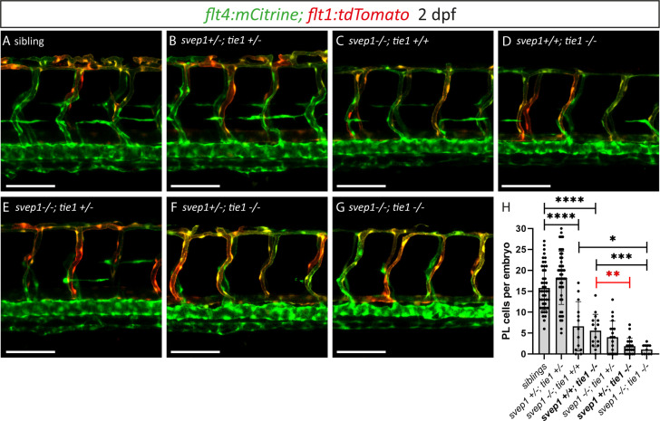
(A–G) Confocal images of blood and lymphatic vasculature in the trunk of 2-dpf-old embryos derived from svep1; tie1 double heterozygous fish, showing severely reduced PL numbers in svep1; tie1 double mutants and significant decrease of PL cell numbers in svep1+/−; tie1−/− compared to svep1+/+; tie1−/− (**p = 0.0012). (H) Quantification of PL cell numbers at 2 dpf using Mann–Whitney test (siblings: n = 45; svep1+/−; tie1+/−: n = 45; svep1−/−; tie1+/+: n = 13; svep1+/+; tie1−/−: n = 15; svep1−/−; tie1+/−: n = 20; svep1+/−; tie1−/−: n = 21; svep1−/−; tie1−/−: n = 11). Scale bar = 100 µm. Values are presented as means ± standard deviation (SD), ****p < 0.0001, ***p = 0.007, *p = 0.0163, ns = not significant. dpf, days post-fertilization; PL, parachordal lymphangioblast.
Heterozygous loss of svep1 exacerbates the PL phenotype in tie1 mutants, indicating genetic interaction between svep1 and tie1.