IMAGE

Fig. 2.

ID
ZDB-IMAGE-230119-17
Source
Figures for Soto et al., 2022
Image
Figure Caption

Fig. 2.

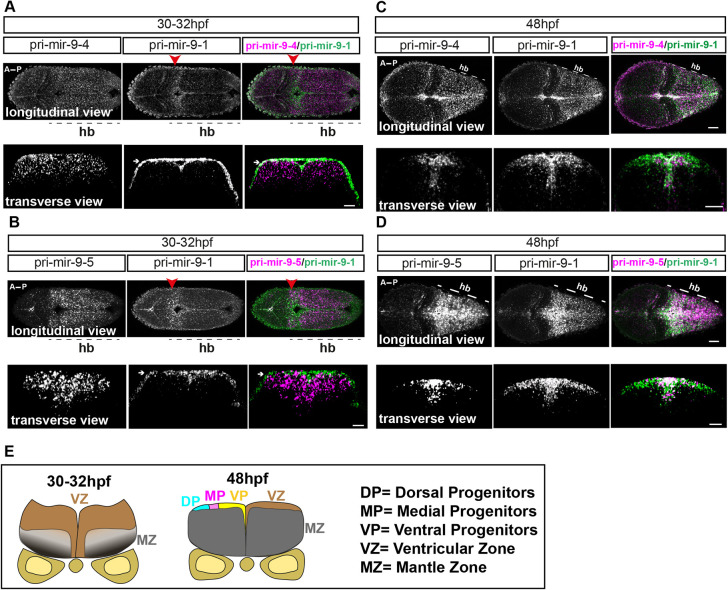
Progressive additive expression of pri-mir-9s during development. (A-D) Representative example of double fluorescent WM-ISH (WM-FISH) labelling of pri-mir-9-1/pri-mir-9-4 (A,C) and pri-mir-9-1/pri-mir-9-5 (B,D) in hindbrain (hb) from wild-type embryo observed at 30-32 hpf (A,B) and at 48 hpf (C,D). Transverse view was collected from hindbrain rhombomere 4/5. Longitudinal view was collected from embryos with anterior to the left and posterior to the right; images are maximum intensity projection; 5 μm thickness for 48 hpf embryos and 10 μm thickness for 30-32 hpf embryos. Merged images indicate pri-mir-9-4 or -9-5 in magenta and pri-mir-9-1 in green. White arrows indicate artefactual signal originated from the amplification step with FITC staining in the WM-FISH; red arrowheads indicate rhombomere 1 of the hindbrain. Pri-mir-9-1/pri-mir-9-4: longitudinal/30-32 hpf, N=3; transverse/30-32 hpf, N=3; longitudinal/48 hpf, N=4; transverse/48 hpf, N=8. Pri-mir-9-1/pri-mir-9-5: longitudinal/30-32 hpf, N=3; transverse/30-32 hpf, N=4; longitudinal/48 hpf, N=4; transverse/48 hpf, N=5. (E) Schematic of transverse section from zebrafish hindbrain at the level of the otic vesicle for 30-32 hpf and 48 hpf. A, anterior; MZ, mantle zone; P, posterior; VZ, ventricular zone. Within the VZ there are dorsal progenitors (DP), medial progenitors (MP) and ventral progenitors (VP). Scale bars: 30 µm.

Acknowledgments
This image is the copyrighted work of the attributed author or publisher, and ZFIN has permission only to display this image to its users. Additional permissions should be obtained from the applicable author or publisher of the image. Full text @ Development