
Image
|
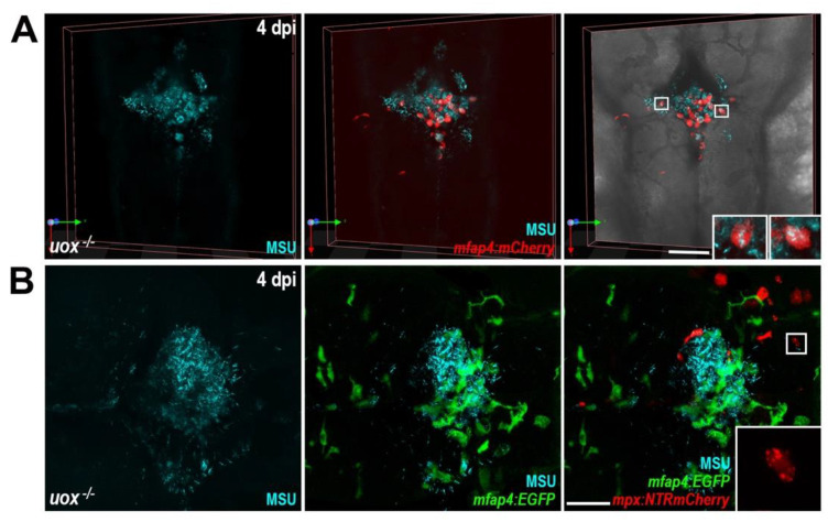
Figure Caption
Figure 6
MSU crystal aggregates within uricase-deficient larvae are dominated by macrophages. (A) Live confocal imaging of MSU crystals within Tg(mfap4:mCherry);uox−/− larvae at 4 days post injection (dpi). Insets, magnified views of boxed regions showing MSU crystal-laden macrophages. (B) Live confocal imaging of MSU crystals within Tg(mfap4:EGFP);(mpx:NTRmCherry);uox−/− larvae at 4 dpi. Inset, magnified view of boxed region showing red fluorescent neutrophil debris. Scale bars: 100 µm in (A), 50 µm in (B).
Figure Data
Acknowledgments
This image is the copyrighted work of the attributed author or publisher, and
ZFIN has permission only to display this image to its users.
Additional permissions should be obtained from the applicable author or publisher of the image.
Full text @ Genes (Basel)