IMAGE

FIGURE 2

ID
ZDB-IMAGE-220923-57
Source
Figures for Talbot et al., 2022
Image
Figure Caption

FIGURE 2

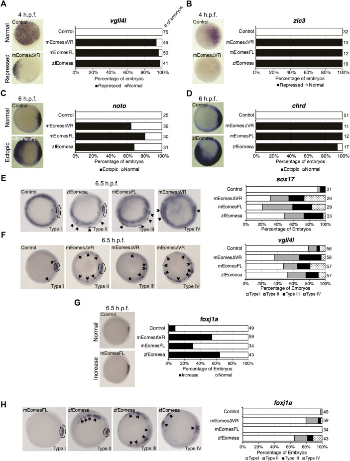
Both FL and ∆VR isoforms of mouse Eomes are functionally equivalent to zebrafish Eomesa in the early embryo. WISH analysis of ectoderm markers vgll4l and zic3 in mid/late blastulas (4 h.p.f.) embryos (A,B), organiser markers noto and chrd(C,D) in early gastrulas (6 h.p.f.), or DFC markers sox17, vgll4l and foxj1a(E–H) in early/mid gastrulas (6.5 h.p.f.). Embryos have been injected at the 1 cell stage to overexpress either mouse EomesFL, Eomes∆VR or zebrafish Eomesa. N = 2. Total numbers of embryos scored per condition are indicated. Representative images of expression patterns per gene per category are shown. (A–D) Animal views; dorsal to the right. (E) Animal views; dorsal to the right. (F–H) Vegetal views; dorsal to the right. Panel G indicates the percentage of embryos with greater intensity of dorsal foxj1a WISH staining, while panel H indicates percentages of embryos with ectopic foxj1a staining. Type I–wild type expression; Type II–ectopic dorsolateral expression with clear primary dorsal DFC cluster; Type III–dorsolateral expression with no defined primary cluster; Type IV–ectopic expression in the ventral margin. Dotted ovals indicate primary DFC clusters. Arrowheads indicate ectopic DFC marker expression.

Acknowledgments
This image is the copyrighted work of the attributed author or publisher, and ZFIN has permission only to display this image to its users. Additional permissions should be obtained from the applicable author or publisher of the image. Full text @ Front Cell Dev Biol