
|
Fig. 2
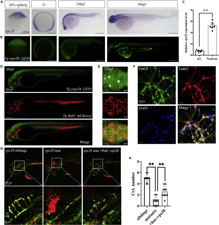
Vps28 was enriched in CNS and cultured mouse cortical neurons
(A) The expression pattern of vps28 at 30% epiboly, 5-somite (5s), 24 hpf, and 36 hpf stage in zebrafish.
(B) Relative expression pattern of vps28 in Tg (Vps28: GFP).
(C) Relative expression level of vps28 in zebrafish ECs and neurons, which were sorted by flow cytometry from Tg (Kdrl: eGFP) and Tg (Huc: eGFP) embryos at 2 dpf, respectively.
(D and E) Expression of vps28 in Tg (vps28: eGFP; Kdrl: mCherry) at 48 hpf and 72 hpf. vps28 was mainly expressed in the zebrafish CNS than DLV (arrowhead) and MsV (arrow) at 72 hpf; DLV, dorsal longitudinal vein; MsV, Mesencephalic vein (E).
(F) Confocal images of primary mouse cortical neurons immunolabeled with Vps28 at 5 days in culture, a majority of the Tubb3 expressing neurons express detectable levels of Vps28 in primary mouse cortical neurons.
(G) Effects of vps28 driven by the HuC promoter (huc: vps28) on CtAs defects of vps28 mutants.
(H) Graphical representations of the CtAs numbers in (G). Data are represented as mean +/− SD. ∗∗p < 0.01.