
|
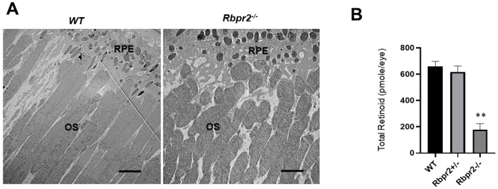
Fig. 5
Rbpr2−/− mice on vitamin A-deficient diets show decreased ocular retinoid concentrations and shorter photoreceptor outer segments. (A) Ultrastructural analysis of photoreceptors using transmission electron microscopy (TEM): Representative TEM images of photoreceptors from 12-week old WT and Rbpr2−/− mice on vitamin A-deficient diets are presented. Scale bar = 600 μm. RPE, retinal pigmented epithelium; OS, outer segments. Data are representative of n = 6 retinal sections per eye from n = 4 mice per genotype. (B) Quantification of total retinoid concentrations (pmole/eye) in eyes from WT, Rbpr2+/−, and Rbpr2−/− mice (eyes from n = 8 mice per genotype). ** p < 0.005, Rbpr2−/− compared to controls.