
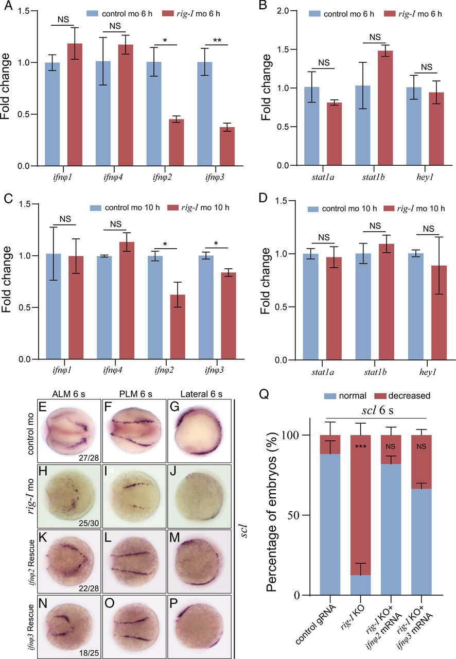
Fig. 7
|
Fig. 7
Examination of the involvement of IFN signaling downstream of RIG-I in the emergence of hematopoietic precursors
(A and B) Quantitative PCR results of ifnφ1-4 at 6 and 10 hpf in control MO or rig-I morphant embryos. (C and D) Quantitative PCR results of stat1a, stat1b, and hey1 at 6 and 10 hpf in control MO or rig-I morphant embryos. (E−P) Expression of hematopoietic precursor marker scl/tal1 at the six-somite stage in embryos injected with control MO from dorsal view (E and F) and lateral view (G), rig-I MO from dorsal view (H and I) and lateral view (J), or rig-I MO and ifnφ2 mRNA from dorsal view (K and L) and lateral view (M), or ifnφ3 mRNA from dorsal view (N and O) and lateral view (P). Images were captured under Olympus stereoscope (MVX10 MacroView; original magnification ×50). (Q) Percent of embryos with phenotype of normal or decreased staining intensity in control or rig-I morphant embryos from (E)–(M) based on WISH analysis. Overexpression of ifnφ2-3 mRNAs partially rescued the defect of hematopoietic precursors in rig-I morphant. Each experiment was repeated three times (n = 3, mean ± SD, Student t test, *p < 0.05, **p < 0.01, ***p < 0.001).