IMAGE

Fig. 5.

ID
ZDB-IMAGE-220304-6
Source
Figures for Allanki et al., 2021
Image
Figure Caption

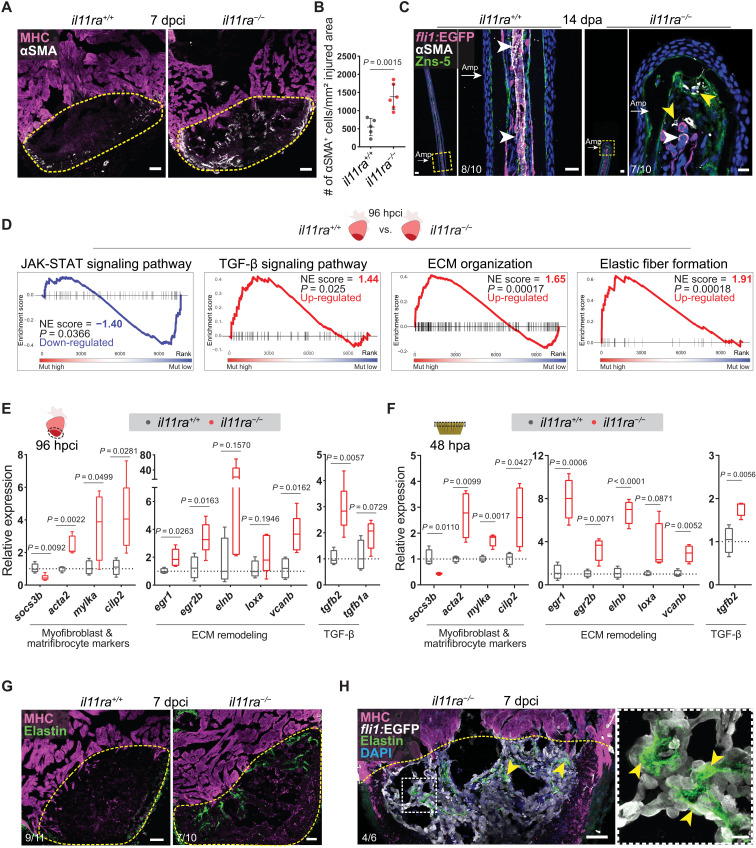
Fig. 5. Il-11 signaling limits myofibroblast differentiation and profibrotic ECM remodeling after injury.

(A) Immunostaining for αSMA (white) and MHC (magenta) expression on cryosections from cardiac ventricles (wt siblings, n = 5; mut, n = 6; 7 dpci). (B) Quantification of αSMA+ cell density. (C) Immunostaining for αSMA (white), GFP (magenta), and Zns-5 antigen (scleroblasts, green) expression on longitudinal cryosections from Tg(fli1:EGFP) caudal fins (wt, n = 10; mut, n = 10; 14 dpa). (D) GSE analysis plots for Reactome and KEGG pathway terms from il11ra−/− versus wild-type sibling adult ventricle transcriptomic analyses, 96 hpci. (E and F) RT-qPCR analysis on dissected injured areas from cardiac ventricles (E) (wt siblings, n = 4; mut, n = 5; 96 hpci) and caudal fins (F) (wt siblings, n = 4; mut, n = 4; 48 hpa) for selected profibrotic gene expression levels. (G and H) Immunostaining [(G) Elastin, green; MHC, magenta; (H) GFP, white] on cryosections from cardiac ventricles [(G) wt siblings, n = 11; mut, n = 10; (H) Tg(fli1:EGFP); mut, n = 6; 7 dpci]. Data represent means ± SD (B), and box plots (E and F) show median, interquartile range (IQR; box margins), and 5th and 95th percentiles (whiskers). Student’s t tests (B, E, and F). n, ventricles (A, E, G, and H); n, caudal fins (C); n, pools of two caudal fins (F). Yellow dashed lines demarcate the injured area (A, G, and H); yellow arrowheads point to αSMA+ myofibroblasts in il11ra mutant fins (C) and Elastin1 expression associated with endocardial cells in the injured area [inset in (H)]; white arrowheads point to vessel-associated αSMA+ smooth muscle cells (C); white arrows point to the amputation plane (C). Ct values are listed in table S5. Scale bars, 50 μm (A, C, G, and H), 20 μm [(C) left inset], and 10 μm [(C) right inset and (H) inset].

Acknowledgments
This image is the copyrighted work of the attributed author or publisher, and ZFIN has permission only to display this image to its users. Additional permissions should be obtained from the applicable author or publisher of the image. Full text @ Sci Adv