IMAGE

FIGURE 1

ID
ZDB-IMAGE-210526-17
Genes
Antibodies
Source
Figures for Grivas et al., 2021
Image
Figure Caption

FIGURE 1

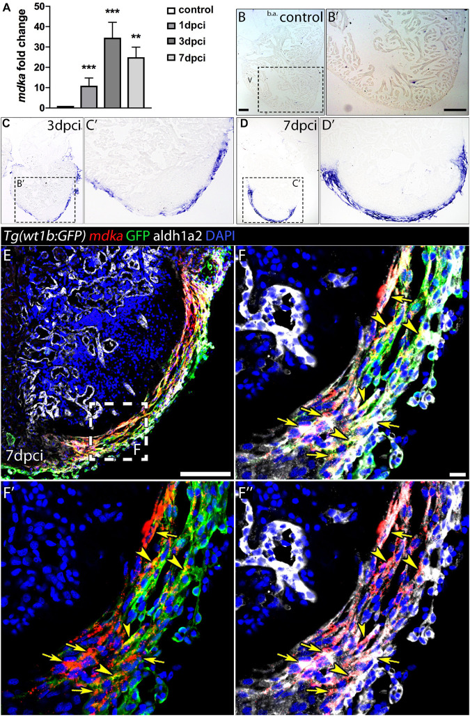
mdka expression upon injury. (A) qPCR analysis of mdka in regenerating hearts. Brown-Forsythe and Welch ANOVA tests; **P < 0.01, ***P < 0.001; Mean ± SD. (B–D′) ISH of mdka in intact (B,B′), 3 dpci (C,C′), and 7 dpci (D,D′) hearts. V, ventricle; b.a., bulbus arteriosus. Scale bars, 100 μm. (E–F′′) Fluorescent ISH for mdka followed by immunolabeling for GFP and Aldh1a2 in 7 dpci Tg(wt1b:GFP) hearts. Arrowheads indicate mdka-GFP overlap and arrows mdka-Aldh1a2 overlap. Scale bar E, 100 μm; (F–F′′), 10 μm.

Figure Data
Acknowledgments
This image is the copyrighted work of the attributed author or publisher, and ZFIN has permission only to display this image to its users. Additional permissions should be obtained from the applicable author or publisher of the image. Full text @ Front Cell Dev Biol