
Fig. 5
- ID
- ZDB-IMAGE-210512-95
- Publication
- Hwang et al., 2021 - Phloroglucinol and dieckol isolated from Ecklonia cava suppress impaired diabetic angiogenesis; A study of in-vitro and in-vivo
- All Figures
- Figures for Hwang et al., 2021
|
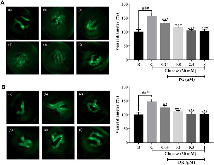
Fig. 5 Inhibitory effects of phloroglucinol (PG) and dieckol (DK) on the increase in retinal vessel diameter in transgenic (flk:EGFP) zebrafish embryos treated with a high concentration of glucose. (A) Fluorescence images of the hyaloid retinal vessels of the transgenic zebrafish (flk:EGFP) embryos treated with PG. (a) 0 mM glucose + 0 µM PG; (b) 130 mM glucose + 0 µM PG; (c) 130 mM glucose + 0.24 µM PG; (d) 130 mM glucose + 0.8 µM PG; (e) 130 mM glucose + 2.4 µM PG; and (f) 130 mM glucose + 8 µM PG; Quantitation of the hyaloid retinal vessel diameter following treatment with PG. (B) Fluorescence images of the hyaloid retinal vessels of transgenic zebrafish (flk:EGFP) embryos treated with DK. (a) 0 mM glucose + 0 µM DK; (b) 130 mM glucose + 0 µM DK; (c) 130 mM glucose + 0.03 µM DK; (d) 130 mM glucose + 0.1 µM DK; (e) 130 mM glucose + 0.3 µM DK; and (f) 130 mM glucose + 1 µM DK; Quantitation of the hyaloid retinal vessel diameter following treatment with DK. The effect of 130 mM of glucose was compared to that of B; blank (0 mM glucose + 0 μM PG, DK), ### p < 0.001. Percentage gap closure was normalized to that of C: control (30 mM glucose + 0 μM PG, DK); ns, not significant, **p < 0.01, *** p < 0.001.