IMAGE

Fig. 4

ID
ZDB-IMAGE-210215-27
Genes
Antibodies
Source
Figures for Yang et al., 2020
Image
Figure Caption

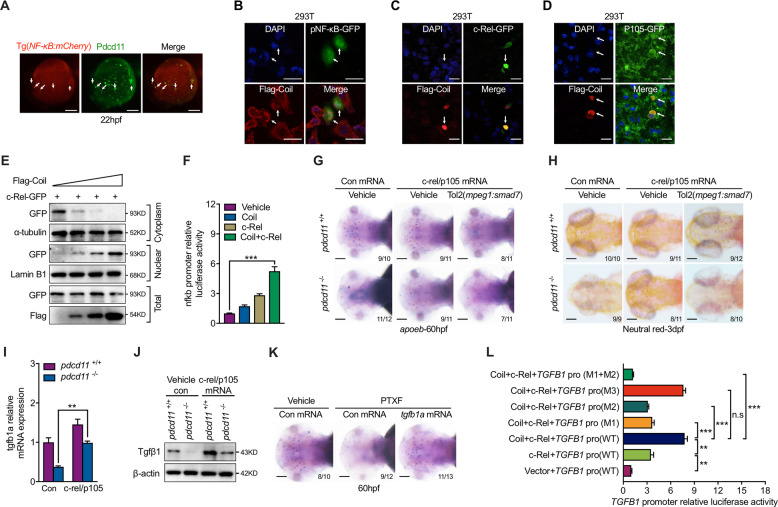
Fig. 4 PDCD11 promotes c-Rel mediated NF-κB activation.

a IF assay of Pdcd11 expression in Tg(NF-κB:mCherry) transgenic line. White arrowheads indicate the colocalized cells on the YS at 22 hpf. Scale bar: 100 μm. b In vitro transfection of plasmid containing NF-κB recognition motif driven GFP and Flag-Coil. White arrows indicate that cells with nuclear Coil expression displayed NF-κB activation. Scale bar: 20 μm. Representative image show cells co-transfected with Flag-Coil and c-Rel (c) or P105 (d). White arrows indicate that cells with nuclear expressed Coil and c-Rel (c) or P105 (d). Scale bar: 20 μm. e. Western blot showing the effect of increasing Coil transfection on nuclear/cytoplasm c-Rel content. f Influence of Coil and c-Rel transfection on luciferase activities driven by the NF-κB-binding site. Microglia numbers in WT and pdcd11 mutants examined by apoeb (g) or Neutral Red staining (h) with c-rel and p105 mRNA or combined mpeg1 promoter driven smad7 plasmid injection. Scale bar: 100 μm. qPCR (i) and western blot (j) assessment of tgfb1a or Tgfβ1 level in 22 hpf WT and pdcd11 mutants with c-Rel and p105 mRNA overexpression. k Microglia numbers in embryos with c-Rel inhibition (PTXF) or combined tgfb1a mRNA complementation. Scale bar: 100 μm. l Reporter assay showing the regulation of TGFB1 promoter by Coil and c-Rel transfection. TGFB1 WT promoter containing three c-Rel-binding sites (5′-ccGGGGcacccccc-3′, −761 bp to TSS; 5′-ggGGGGacgccccgt-3′, −776 bp to TSS; 5′-aaGGGAcccccctcg-3′, −1019 bp to TSS). The three binding motifs were respectively mutated and designated as M1 (5′-ccAAAAcacccccc-3′, −761 bp to TSS), M2 (5′-ggAAAAacgccccgt-3′, −776 bp to TSS), M3 (5′-aaAAACcccccctcg-3′, −1019 bp to TSS). The number positioned in the lower right corner of g, h, k represents the number of zebrafish embryos shown positive phenotypes versus the total number of embryos examined. Means ± SEM are shown for three independent experiments. n.s not significant, *P < 0.05; **P < 0.01; ***P < 0.001 (Student’s t test).

Figure Data
Acknowledgments
This image is the copyrighted work of the attributed author or publisher, and ZFIN has permission only to display this image to its users. Additional permissions should be obtained from the applicable author or publisher of the image. Full text @ Cell Death Differ.