
|
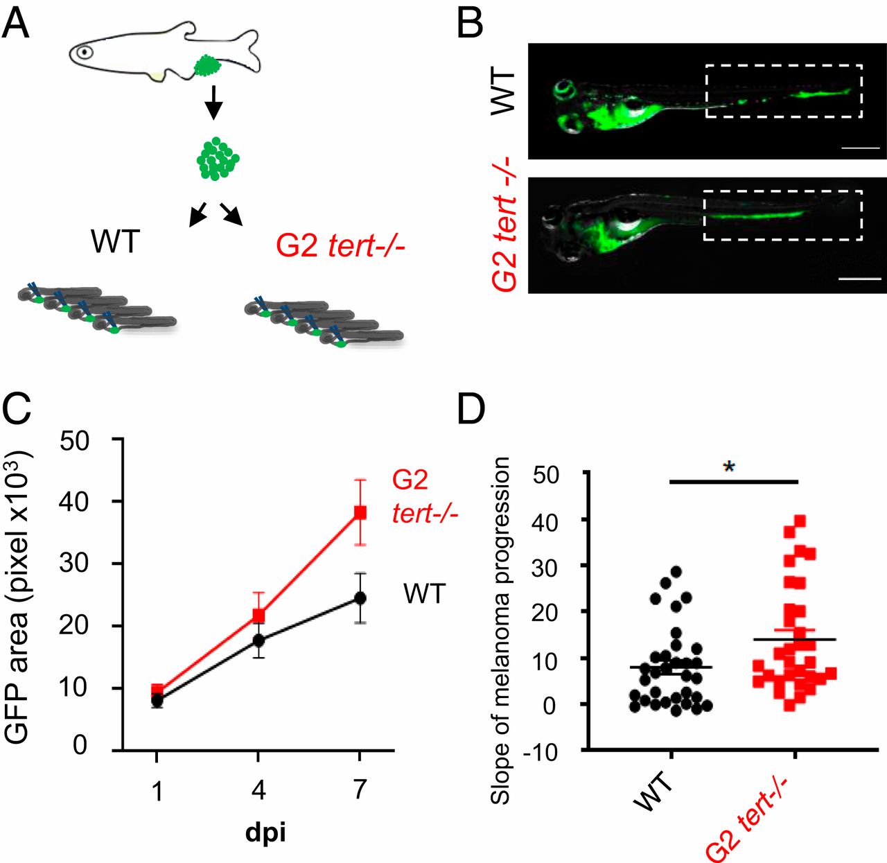
Fig. 3 G2 tert−/− larvae with very short telomeres exhibit increased melanoma expansion. (A) Experimental design for melanoma allotransplants in zebrafish larvae. Melanoma tumors were dissected from Tg(mitfa:HRASG12V; β-actin:GFP) zebrafish and cells dissociated. HRAS melanoma cells were then injected into blood circulation of 2-dpf zebrafish larvae. Larvae were kept for 7-d-postinjection (7-dpi). (B) Representative images of HRAS melanoma cells spread (green) in WT or G2 tert−/− larvae at 7 dpi. (Scale bars: 0.5 mm.) (C) Time course of HRAS melanoma cells spread in a group of WT and G2 tert−/− larvae (P < 0.01 at 7 dpi; WT n = 10; G2 tert−/− n = 11). (D) Melanoma tumors are more disseminated in G2 tert−/− larvae (*P < 0.05; WT n = 32; G2 tert−/− n = 31). A linear regression of three time points (1, 4, and 7 dpi) was used to calculate the slope of melanoma expansion over time. Each dot represents one larvae allotransplant.