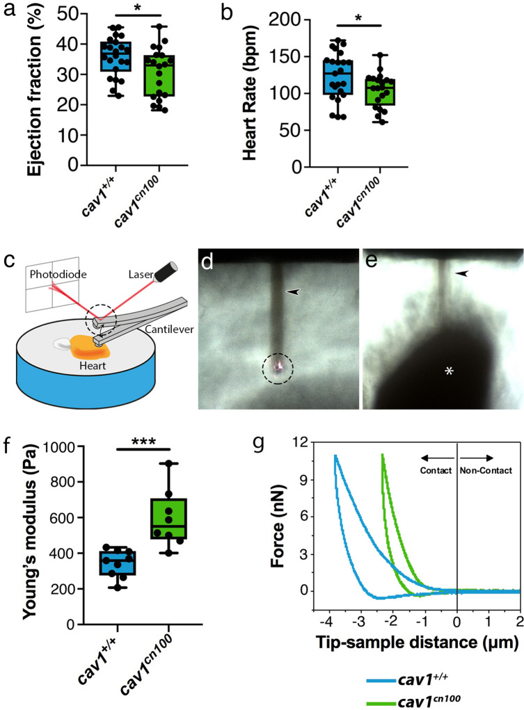
IMAGE

Figure 6

ID
ZDB-IMAGE-200805-13
Source
Figures for Grivas et al., 2020
Image
Figure Caption

Figure 6

Impaired cardiac function and stiffer heart tissue in caveolae-deprived cav1cn100 mutants. (a) Quantification of ejection fraction in cav1+/+ and cav1cn100 hearts. nWT = 22, ncn100 = 20. (b) Heart rate measurements in cav1+/+ and cav1cn100 animals. nWT = 23, ncn100 = 20. t-test, *P < 0.05. (c) AFM set-up. Cartoon made with Adobe Illustrator CC2018 (www.adobe.com). (d, e) Images of the cantilever (arrowhead), the laser beam on top of the cantilever (dashed circle, also in c) and the ventricle (asterisk). Images were taken with an inverted optical microscope (AXIO Observer D1; Carl Zeiss, Germany, see Methods). (f) Biomechanical characterization of cav1+/+ and cav1cn100 ventricles as measured by AFM-force spectroscopy and expressed in Young’s moduli. nWT = 9, ncn100 = 8, t-test, ***P < 0.001. (g) Force-distance graph for indentation and retraction of the cantilever over the ventricular surface.

Figure Data
Acknowledgments
This image is the copyrighted work of the attributed author or publisher, and ZFIN has permission only to display this image to its users. Additional permissions should be obtained from the applicable author or publisher of the image. Full text @ Sci. Rep.