IMAGE

Figure 2

ID
ZDB-IMAGE-200627-3
Genes
Source
Figures for Weaver et al., 2020
Image
Figure Caption

Figure 2

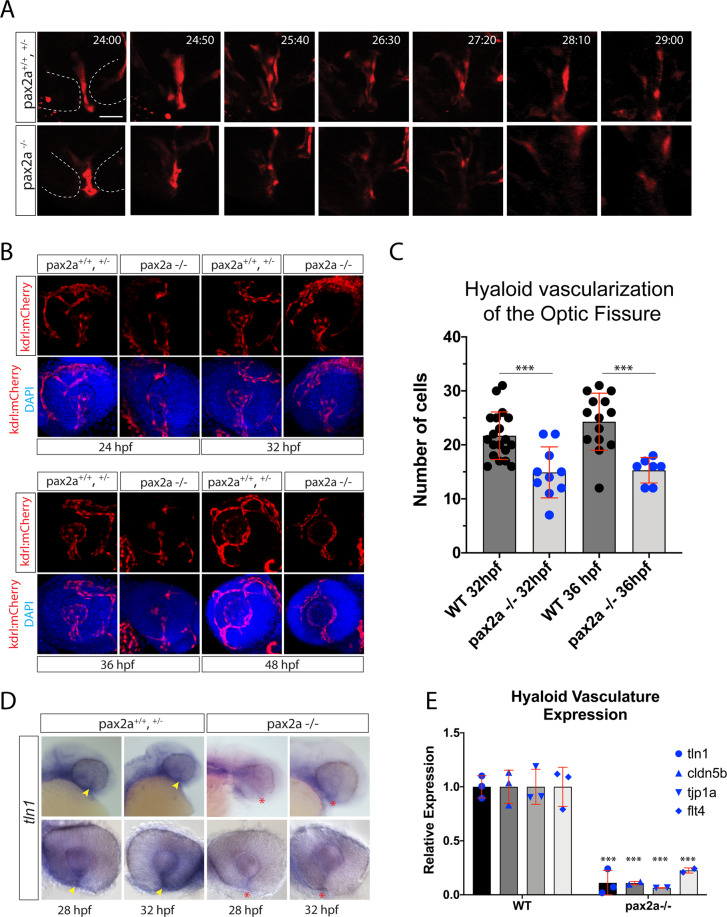
Pax2a is necessary for recruitment of hyaloid vasculature into the optic fissure. (A)In vivo 4D confocal imaging of Tg[kdrl:mCherry] pax2a+/+,+/− or pax2a−/− embryos. Time lapse series depicting the region of the OF (dotted white lines) and mCherry positive vasculature endothelial cells migrating through the fissure from 24-29 hpf. Scale bar = 10 μm. (B) Comparison of pax2a+/+,+/− and pax2a−/− vascularization during OF fusion, 24-48 hpf. 3D reconstructions of whole mount Tg[kdrl:mCherry] (red) pax2a+/+,+/− or pax2a−/− embryos with DAPI (blue) stained DNA. Scale bar = 50 μm. (C) Quantification of the number of mCherry positive cells from 3D confocal stacks within the region of the OF. Individual embryo results are depicted. ANOVA p < 0.0001. (D) Whole mount in situ hybridization comparing tln1 expression between pax2a+/+,+/− and pax2a−/− embryos at 28 and 32 hpf. tln1 signal within the OF (yellow arrowhead) appears reduced in Pax2a−/− embryos (red *). (E) qPCR analysis of tln1 expression from heads of pax2a+/+ and pax2a−/− embryos at 32 hpf confirms a reduction in tln1 expression.

Figure Data
Acknowledgments
This image is the copyrighted work of the attributed author or publisher, and ZFIN has permission only to display this image to its users. Additional permissions should be obtained from the applicable author or publisher of the image. Full text @ Sci. Rep.