
|
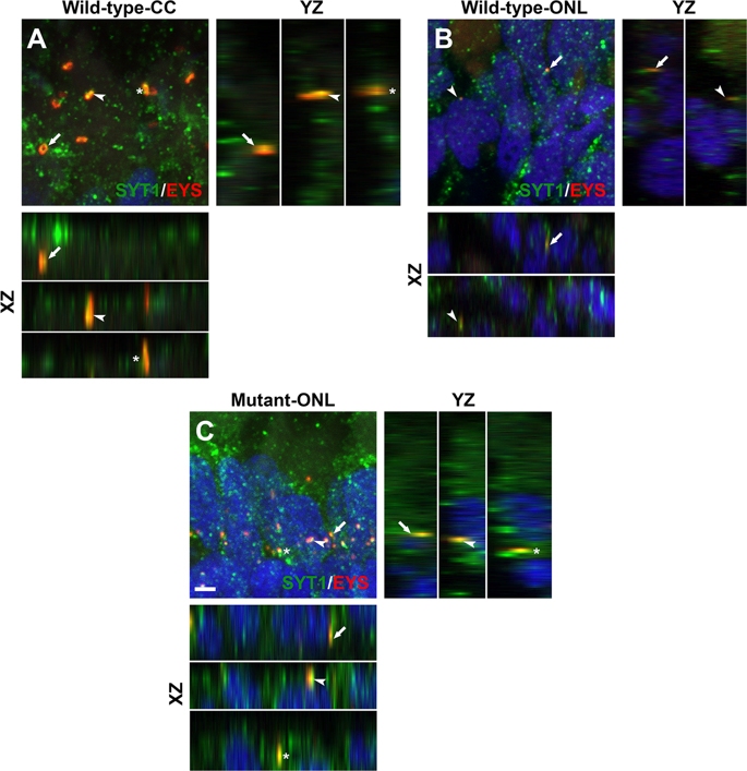
Fig. 4
Mislocalized EYS in pomgnt1 mutant zebrafish retina was co-localized with synaptogamin-1. Retinal sections from 2-mpf zebrafish were double stained with antibodies against EYS (red fluorescence) and synaptotagmin-1 (green fluorescence). (A) Wild-type inner/outer segment layers showing connecting cilia (CC). Maximal projection and its orthogonal views of three double stained puncta are shown. EYS-immunoreactivity was co-localized with synaptagmin-1 reactivity. (B) Wild-type outer nuclear layer (ONL). Maximal projection and its orthogonal views of two double stained puncta are shown. EYS-immunoreactivity was co-localized with synaptagmin-1 reactivity. (C) Homozygous pomgnt1sny7 mutant outer nuclear layer. Maximal projection and its orthogonal views of three double stained puncta are shown. Mislocalized EYS immunoreactive puncta was co-localized with synaptotagmin-1 reactivity. Scale bar in C: 2 µm.