IMAGE

FIGURE 4

ID
ZDB-IMAGE-200423-20
Genes
Antibodies
Source
Figures for Carter et al., 2020
Image
Figure Caption

FIGURE 4

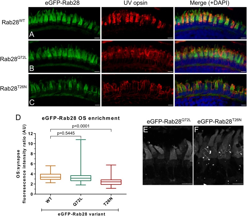
eGFP-Rab28 localization to larval zebrafish cone outer segments is partially dependent on GTP/GDP-binding. (A–C) Representative confocal z-projections of eGFP-Rab28 localization in 5 dpf zebrafish cone photoreceptors. The WT, putative GTP-preferring (Q72L) and GDP-preferring (T26N) variants of Rab28 all localize strongly to the outer segments of zebrafish cones, co-localizing with UV opsin labeling. Scale bars 5 μm. For WT, Q72L and T26N eGFP-Rab28 reporters a total of 14, 24 and 25 larvae were imaged, respectively. (D) Box and whisker plots of the ratio of eGFP-Rab28 intensity in the OS vs. synaptic region of larval cones. Box extremities represent 1st and 3rd quartiles; whiskers are maximum and minimum values. Data are from 60 cones per transgenic line. One-way ANOVA p-value < 0.0001. (E,F) Deconvolved, high resolution confocal z-projections of eGFP-Rab28 Q72L and T26N mutant localization in cones of 5 dpf larvae. A discrete localization pattern of the T26N mutant in COS is clearly observed (white arrowheads). Scale bars 4 μm.

Figure Data
Acknowledgments
This image is the copyrighted work of the attributed author or publisher, and ZFIN has permission only to display this image to its users. Additional permissions should be obtained from the applicable author or publisher of the image. Full text @ Front Cell Dev Biol