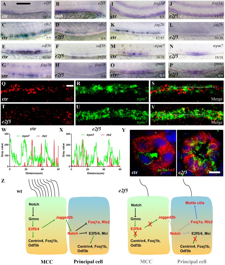
IMAGE

Fig 6

ID
ZDB-IMAGE-200412-15
Genes
Source
Figures for Xie et al., 2020
Image
Figure Caption

Fig 6 Ectopic motile cilia developed in the principal cells of <italic>e2f5</italic> mutants.

(A-P) Whole-mount in situ hybridization results showing the expression of e2f5 and other marker genes in the pronephric duct of 24 hpf control and mutant larvae as indicated. The numbers in the bottom right-hand corners indicate the numbers of embryos with similar staining results (left) and total numbers of embryos analyzed (right). (Q-V) Fluorescence in situ hybridization results showing the expression of rfx2 (red) and trpm7 (green) in the pronephric tubule of 36 hpf wild-type control (Q-S) and e2f5 mutant larvae (T-V). (W-X) Line profile plots showing the pixel intensities of green and red channels along the dotted line in panels S and V. (Y) Immunofluorescence results showing the staining of anti-acetylated tubulin (ac-Tu) and α6F antibodies on cross-sections through the pronephric tubules of 5dpf control and e2f5 mutant larvae. (Z) Model illustrating dual roles of E2f5 during multiciliogenesis. In the absence of E2f5 (maybe also E2f4), both MCC and principal progenitor cells developed a single motile cilium. Scale bars: 500 μm in panels A-P, 10 μm in panels Q-V and 5 μm in panel Y.

Figure Data
Acknowledgments
This image is the copyrighted work of the attributed author or publisher, and ZFIN has permission only to display this image to its users. Additional permissions should be obtained from the applicable author or publisher of the image. Full text @ PLoS Genet.