
Fig. 3
|
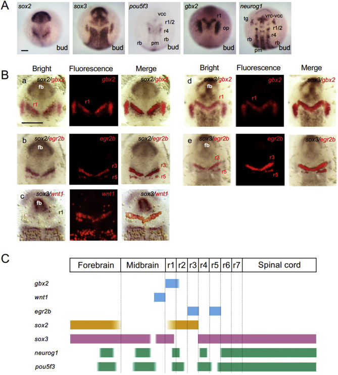
Fig. 3 Localization of the proneural clusters and soxB1 regions in the neural plate. A. Expression of soxB1, pou5f3, gbx2, and neurog1 at the bud stage revealed by single-color WISH. B. Comparison of the gene expression patterns by two-color WISH. The expression domains of two specified genes are stained brown and red. The bright-field views (left), epifluorescence views (middle), and merged views (right) are shown for each of the gene pairs examined. fb, forebrain. For the other abbreviations, see the legend for Fig. 1. Scale bars, 100 μm. C. Schematic views of the expression domains of brain regional marker genes, soxB1, and neurog1/pou5f3.
Reprinted from Developmental Biology, 457(1), Inomata, C., Yuikawa, T., Nakayama-Sadakiyo, Y., Kobayashi, K., Ikeda, M., Chiba, M., Konishi, C., Ishioka, A., Tsuda, S., Yamasu, K., Involvement of an Oct4-related PouV gene, pou5f3/pou2, in neurogenesis in the early neural plate of zebrafish embryos, 30-42, Copyright (2019) with permission from Elsevier. Full text @ Dev. Biol.