
Image
|
Figure Caption
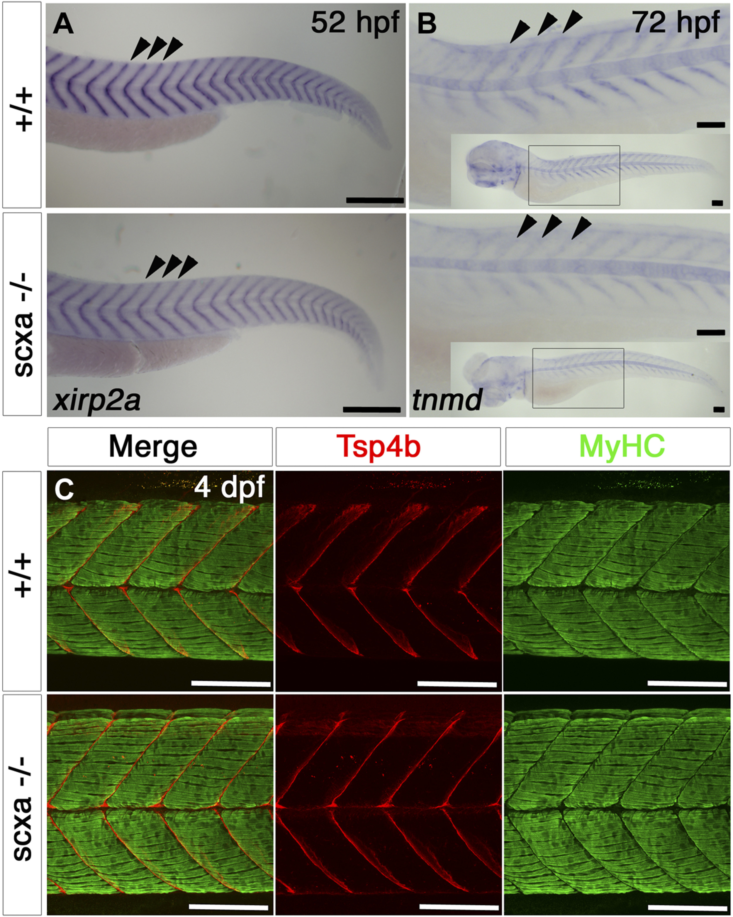
Fig. 6
Scxa mutants have reduced levels of tendon markers, but somitic MTJs appear normal. A, B) In situ hybridization for xirp2a at 52 hpf (A) and tnmd at 3 dpf (B) for scxa−/− and their siblings (+/+), lateral view, anterior to left. B) The main image shows the anterior somites, inset-whole embryo. Both xirp2a and tnmd mRNA levels are reduced at the MTJs at the somitic borders (arrowheads). C) Confocal stacks of immunodetection of MyHC (A4.1025) and Tsp4b in somites 11–14 of 4 dpf embryos of scxa−/− and siblings showing normal distribution of Tsp4b and normal muscle structure. All scale bars, 100 µm.
Figure Data
Acknowledgments
This image is the copyrighted work of the attributed author or publisher, and
ZFIN has permission only to display this image to its users.
Additional permissions should be obtained from the applicable author or publisher of the image.
Full text @ FASEB J.