
Fig. 1
- ID
- ZDB-IMAGE-190816-21
- Genes
- Publication
- Rho et al., 2019 - Rap1b Promotes Notch-Signal-Mediated Hematopoietic Stem Cell Development by Enhancing Integrin-Mediated Cell Adhesion
- All Figures
- Figures for Rho et al., 2019
|
Fig. 1
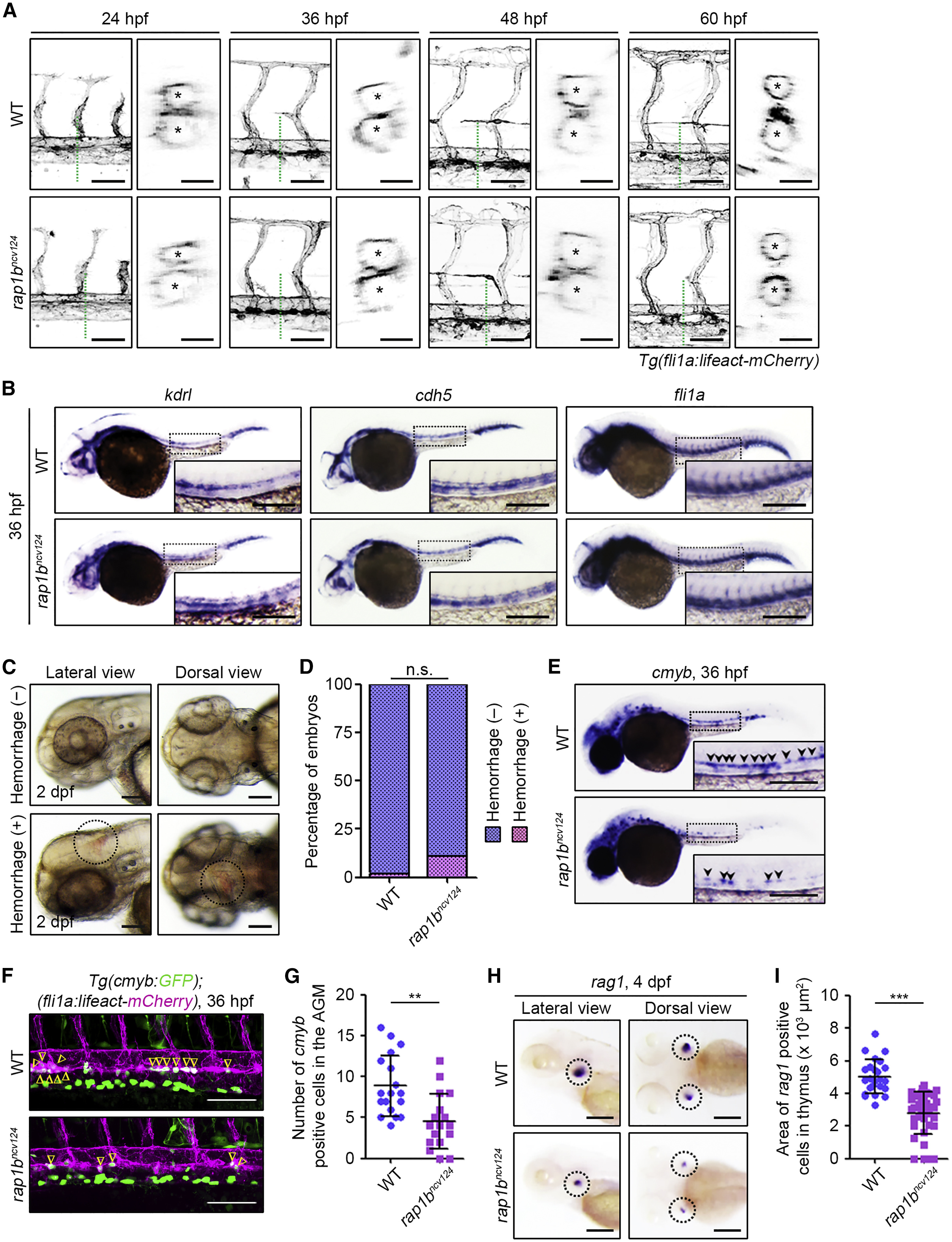
Rap1b Is Not Essential for Vascular Development and Integrity but Is Required for HSC Development
(A) Confocal stack fluorescence images of trunk vasculature in sibling (WT; upper) and rap1bncv124 (lower) embryos and larvae at 24, 36, 48, and 60 hpf with the Tg(fli1a:lifeact-mCherry) background. Lateral views with anterior to the left. The cross-sectional images of the area indicated by dotted green lines on the images are shown on the right side. Asterisks indicate lumens of DA (upper) and posterior cardinal vein (lower).
(B) Expression patterns of endothelial marker genes kdrl (left column), cdh5 (middle column), and fli1a (right column) in sibling (WT; upper) and rap1bncv124 (lower) embryos at 36 hpf.
(C) Representative bright field images of the heads of 2 dpf rap1bncv124embryos without (upper panels) or with (lower panels) intracranial hemorrhage. Lateral and dorsal view images are shown in the left and right columns, respectively. Dotted circles indicate the region of cranial hemorrhage.
(D) Quantification of intracranial hemorrhage in sibling (WT) and rap1bncv124 embryos at 2 dpf. Percentages of normal embryos (blue) and those showing intracranial hemorrhage (pink). WT, n = 69; rap1bncv124, n = 88. n.s., not significant.
(E) Expression patterns of the HSC marker cmyb in 36 hpf sibling (WT) and rap1bncv124 embryos. Arrowheads indicate cmyb-positive HSCs.
(F) Confocal stack fluorescence images of trunk axial vasculature in 36 hpf sibling (WT; upper) and rap1bncv124 (lower) embryos with the Tg(cmyb:GFP);(fli1a:lifeact-mCherry) background. Lateral views with anterior to the left. Merged images of cmyb:GFP (green) and fli1a:lifeact-mCherry (magenta). Arrowheads indicate cmyb:GFP- and fli1a:lifeact-mCherry double-positive HSCs located in the ventral side of the DA.
(G) Number of cmyb:GFP- and fli1a:lifeact-mCherry double-positive HSCs in the aorta-gonad-mesonephros (AGM) regions of 36 hpf sibling (WT) and rap1bncv124 embryos, as observed in (F). Each dot represents an individual embryo. Error bars indicate means ± SD. WT, n = 18; rap1bncv124, n = 17. ∗∗p < 0.01.
(H) Expression patterns of the lymphoid lineage marker rag1 in the 4 dpf sibling (WT) and rap1bncv124 embryos. Lateral (left column) and dorsal (right column) views with anterior to the left. Dotted circles indicate the region of the thymus.
(I) Quantification of the rag1-positive thymus area in the 4 dpf sibling (WT) and rap1bncv124 embryos, as observed in (H). Each dot represents an individual thymus. Error bars indicate means ± SD. WT, n = 12; rap1bncv124, n = 19. ∗∗∗p < 0.001.
In (B) and (E), boxed areas are enlarged in the insets. Scale bars: 50 μm (lateral view in A); 20 μm (cross-sectional image in A); 200 μm (B, C, E, and H); 100 μm (F).
Reprinted from Developmental Cell, 49(5), Rho, S.S., Kobayashi, I., Oguri-Nakamura, E., Ando, K., Fujiwara, M., Kamimura, N., Hirata, H., Iida, A., Iwai, Y., Mochizuki, N., Fukuhara, S., Rap1b Promotes Notch-Signal-Mediated Hematopoietic Stem Cell Development by Enhancing Integrin-Mediated Cell Adhesion, 681-696.e6, Copyright (2019) with permission from Elsevier. Full text @ Dev. Cell